Author: edu

AUTHORS
Carlos S. Hernández
Andrea Gil
Amal Zaytouny
Ignacio Casares
Jesús Poderoso
Alfonso de Lara
Alec Wehse
Shivang R. Dave
Daryl Lim
Eduardo Lage
Nicolas Alejandre
 
JOURNAL Ophthalmic and Physiological Optics
ABSTRACT

Abstract

Introduction

Despite the well-known reproducibility issues of subjective refraction, most studies evaluating autorefractors compared differences between the device and subjective refraction. This work evaluated the performance of a novel handheld Hartmann–Shack-based autorefractor using an alternative protocol, which considered the inherent variability of subjective refraction.

Methods

Participants underwent an initial measurement with a desktop autorefractor, two subjective refractions (SR1 and SR2) and a final measurement with the QuickSee Free (QSFree) portable autorefractor. Autorefractor performance was evaluated by comparing the differences between the QSFree and each of the subjective refractions with the difference between the subjective refractions (SR1 vs. SR2) using Bland–Altman analysis and percentage of agreement.

Results

A total of 75 subjects (53 ± 14 years) were enrolled in the study. The average difference in the absolute spherical equivalent (M) between the QSFree and the SR1 and SR2 was ±0.24 and ±0.02 D, respectively, that is, very similar or smaller than the SR1 versus SR2 difference (±0.26 D). Average differences in astigmatic components were found to be negligible. The results demonstrate that differences between QSFree and both subjective refractions in J0 and J45 were within ±0.50 D for at least 96% of the measurements. The limits of agreement (LOAs) of the differences between QSFree and SR1, as well as QSFree and SR2, were higher than those observed between SR1 and SR2 for MJ0 and J45.

Conclusions

A protocol was designed and validated for the evaluation of a refractive device to account for the variability of subjective refraction. This protocol was used to evaluate a novel portable autorefractor and observed a smaller difference between the device and subjective refractions than the difference between the two subjective refraction measurements in terms of mean bias error, although the standard deviation was higher.


LINK here

AUTHORS
Andrea Gil Ruiz
Carlos Santiago Hernandez Torres
Ignacio Casares
Jesus Poderoso
Daryl Lim
Shivang Dave
Eduardo Lage Negro
JOURNAL ARVO
ABSTRACT

Purpose

Modern autorefractors have demonstrated high accuracy and higher repeatability than manifest refraction in adults yet are not considered precise enough to substitute the gold standard, even in low-resource settings where there is a severe shortage of experienced refractionists. This work evaluates a novel approach that combines dynamic wavefront aberrometry data, acquired using an affordable portable autorefractor, with the analysis of retinal image quality metrics (IQMs) to predict subjective refraction.

Methods

56 subjects (34±14 years) were recruited for the analysis. Each participant underwent standard clinical refraction followed by a 10-second video acquisition using a QuickSee (QS) wavefront aberrometer (PlenOptika, USA). Shack-Hartmann images of each video for the right eye were processed to obtain Zernike coefficients up to the 4th order. Coefficients obtained for each image were mathematically corrected with the closest sphero-cylindrical correction and the residual wavefront error was used to calculate the Point Spread Function (PSF) and IQMs (e.g., Strehl Ratio) describing some performance parameter of the corrected eye. Since each IQM is part of a dynamic sequence, it is possible to build a dynamic signal for each metric which contains information about fluctuations in image quality during the measurement. The final refraction is obtained as the average of the refractions corresponding to the images whose IMQs provided optimal performance.
To assess this method, we compared the results of the QS with those obtained by the new algorithm (using 6 different IQMs, Table 1) against the standard clinical refraction. Figures of merit were mean bias error (MBE), 95% limits of agreement (LOA), mean absolute error (MAE), and the percentages of agreement (0.25 and 0.5D thresholds) between refractions.

Results

The proposed method reduced the differences between QS and manifest refraction. Specifically, for spherical equivalent refraction (M) (Table 1) a 79% average reduction in MBE together with moderate average improvements for MAE (10.3%), LOA (5%), and percentage of agreement within 0.25D (8.8%) and 0.5D (6.3%) were found for all IQMs evaluated.

Conclusions

The proposed algorithm behaves as an efficient filter which selects those measurements within the dynamic sequence that are more representative of the manifest refraction of the patient.

LINK here

AUTHORS
Eduardo Lage
Carlos Santiago Hernandez
Andrea Gil Ruiz
Ignacio Casares
Jesus Poderoso
Alec Wehse
Alfonso de Lara
Daryl Lim
Shivang Dave
JOURNAL ARVO
ABSTRACT

Purpose

High repeatability portable autorefractors may enable effective deploy of global-health initiatives to mitigate uncorrected refractive error worldwide. For example, in studies to evaluate the effectiveness of different measures, repeatability has a direct impact on the statistical power and sample size required, while in screening applications, contributes to increased sensitivity and specificity. This work presents a preliminary assessment of the inter- and intrasession repeatability of an affordable autorefractor (QuickSee, PlenOptika Inc) under two separate measurement conditions, with and without fogging lenses.

Methods

6 volunteers (29,8 ± 8,1 years old) were measured at 2 different sessions spaced 1-week. Each subject was tested 6 times per session with the QuickSee, 3 times in standard mode and 3 using a modified version of the device eyecup to allow over-refraction through fogging lenses (+2.0 D). Intersession measurements were spaced 5 minutes apart and the complete alignment procedure was repeated for each measurement. After converting autorefractor readings into power vectors (M, J0, J45), repeatability was evaluated using the Bland-Altman method to compare differences between all possible combinations from each subject (inter- and intrasession). Repeatability coefficients (RPC), estimated as the 95% limits of agreement, and the mean absolute errors were also evaluated. In all cases, only results for the right eyes were analyzed.

Results

Participants had an average Spherical Equivalent (SE) refraction of -0.75 ± 2.06 D, (Min -5.11 D, Max 0.175 D). A total of 36 samples per dataset were obtained for the intrasession analysis while 54 samples per dataset were used in the intersession comparison. In all cases the mean difference was practically 0 indicating no bias for any of the vector components. SE RPCs for intrasession test were 0.15 D in standard mode and 0.29 D with the fogging lenses. Intersession RPCs were 0.29 D and 0.4 D without and with the fogging lenses, respectively. Detailed results for all vector components are shown in Table 1.

Conclusions

This preliminary analysis shows that the device can provide excellent RPCs in standard mode, which decrease moderately with the use of the fogging lenses. Further work exploring improvements in the eyecup design and measurement averaging techniques may further enhance the RPC values in over-refraction mode.

LINK here

AUTHORS
Andrea Gil
Carlos S. Hernandez 
Ahhyun Stephanie Nam
Varshini Varadaraj
Nicholas J. Durr
Daryl Lim
Shivang R. Dave
Eduardo Lage
JOURNAL Scientific Reports
ABSTRACT

The aim of this work is to evaluate the performance of a novel algorithm that combines dynamic wavefront aberrometry data and descriptors of the retinal image quality from objective autorefractor  measurements to predict subjective refraction. We conducted a retrospective study of the prediction accuracy and precision of the novel algorithm compared to standard search-based retinal image quality optimization algorithms. Dynamic measurements from 34 adult patients were taken with a handheld wavefront autorefractor and static data was obtained with a high-end desktop wavefront aberrometer. The search-based algorithms did not significantly improve the results of the desktop system, while the dynamic approach was able to simultaneously reduce the standard deviation (up to a 15% for reduction of spherical equivalent power) and the mean bias error of the predictions (up to 80% reduction of spherical equivalent power) for the handheld aberrometer. These results suggest that dynamic retinal image analysis can substantially improve the accuracy and precision of the portable wavefront autorefractor relative to subjective refraction.

LINK here

Funding Agency Translational Research Institute for Space Health-NASA
Principal Investigator/s Shivang R. Dave
Information Type of Grant: Competitive funding , United states

Grant code: NASA NNX16AO69A

Awardee Organization: Plenoptika Inc

Project Start Date: 29-November-2021

Project End Date: 29-November-2022

Total Funding: $90,000

MEDIC Personnel included in the grant / Role:

Dr. Eduardo Lage — Investigator

MsC Carlos Hernandez – Investigator

MsC Andrea Gil — Investigator

Continue reading

AUTHORS
Sanil Joseph, MHA, MSc
Varshini Varadaraj, MD, MPH
Shivang R. Dave, PhD
Eduardo Lage, PhD
Daryl Lim, PhD
Kanza Aziz, MD
Sarah Dudgeon, (MPH)
Thulasiraj D. Ravilla, MBA
David S. Friedman, MD, PhD, MPH 
JOURNAL Ophthalmology
ABSTRACT

Abstract

Purpose

To compare patient preferences for eye glasses prescribed using a low-cost portable wavefront autorefractor versus standard subjective refraction.

Design

Randomized, cross-over clinical trial

Participants

Patients aged 18-40 years presenting with refractive errors to a tertiary eye hospital in Southern India.

Methods

Participants underwent subjective refraction (SR) followed by autorefraction (AR) using the monocular version of the QuickSee device. An independent optician, masked to the refraction approach, prepared eyeglasses based on each refraction approach. Participants (masked-to-refraction source) were randomly assigned to use either SR- or AR-based eyeglasses first, followed by the other pair, for a week each. At the end of each week, participants had vision checked and were interviewed about their experience with the eyeglasses.

Main outcome measures

Proportion of patients preferring eyeglasses prescribed using AR and SR

Results

The 400 participants enrolled between March 26, 2018 and August 02, 2019, had mean (SD) age of 28.4 (6.6) years and 68.8% were women. There was strong correlation between spherical equivalents using SR and AR (r=0.97, p<0.001) with a mean difference of -0.07 diopters (95% limits of agreement: -0.68 to 0.83). Of the 301 (75.2%) patients who completed both follow-up visits, 50.5% (n=152) and 49.5% (n=149) preferred glasses prescribed using SR and AR, respectively (95% CI: 45.7% – 56.3%; p=0.86). There were no differences in demographic or vision characteristics between participants with different preferences (p>0.05 for all).

Conclusions

We observed strong agreement between the prescriptions from SR and QR, and eyeglasses prescribed using SR and AR were equally preferred by patients. Wider use of prescribing based on AR alone in resource-limited settings is supported by these findings.
LINK here

During the last 25th of Febraury, the event “Learn about the best TFG of the 2019-20 academic year and how Deloitte participates in innovative and entrepreneurial projects” took place in the Autonomous University of Madrid. This time online, as a result of the current situation. Among other attendees, representing the UAM was Mr. Javier Ortega García, Vice-rector of Innovation, Transfer and Technology, who made the introduction to the event, and from Deloitte, Mr. Francisco Faraco, Partner at Deloitte – Fraud Prevention, who gave the award to the winner of the best bachelor thesis at the E.P.S. during the 2019-20 academic year, enrolled in Cybersecurity, Data Science and Artificial Intelligence categories. As it was said in the event ‘At Deloitte we reward talent and innovation with initiatives of this type’.

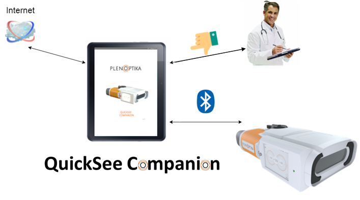
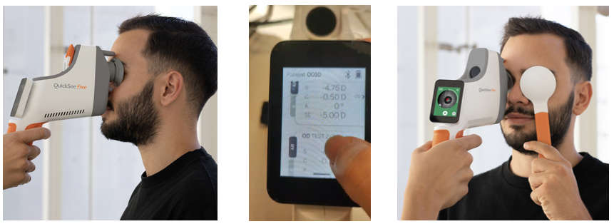
In this occasion the awarded was our engineer Ignacio Salinas Sánchez for his work “Development of a Calibration system for a Visual Simulation device”. The medical device was the SimVis developed by 2eyesVision: a system that prescribes multifocal lenses allowing patients to know if they adapt correctly to their needs before to surgical implantation.

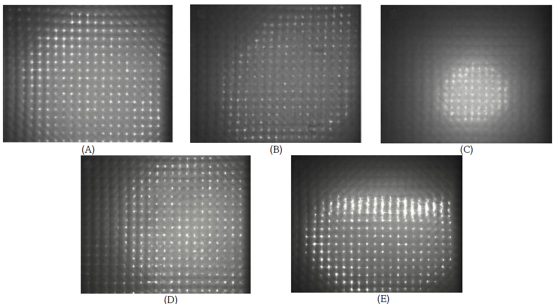
The project consists not only of a software part that includes three main modules: camera, communication and image processing but also a hardware prototype with the function of carrying out verification and validation tests.

“It has been a unique opportunity and a pleasure to collaborate in the development of medical devices with the objective of helping other people, which gives you an added bonus of desire, energy and enthusiasm”, were some of Ignacio’s words.

The Jury also awarded some Special Mentions with their corresponding Diploma of Recognition to Alberto Ruiz Guijosa, Marta Simón Pinacho and Diego Sainz de Medrano Otero, for the quality of their works submitted to the contest.

In addition, during the session other topics were discussed such as Smart Stadiums, Gaming projects, “a life experience” by Álvaro Pérez (Senior Analytical Consultant at Google) and finally a success story from the entrepreneurship office of the UAM – UAM Emprende.

Thank both, Deloitte and UAM for giving us the opportunity to participate in this kind of innitiatives, where all the work, effort and time invested in these projects is recognized.

AUTHORS
Andrea Gil
Carlos S. Hernández
Pablo Pérez-Merino
Marcos Rubio
Gonzalo Velarde
María Abellanas-Lodares
Ángeles Román-Daza
Nicolás Alejandre
Ignacio Jiménez-Alfaro
Ignacio Casares
Shivang R. Dave
Daryl Lim
Eduardo Lage
JOURNAL PLoS ONE 15(10): e0240933
ABSTRACT

ABSTRACT

Purpose
To assess the performance of an open-view binocular handheld aberrometer (QuickSee) for diagnosing refractive errors in children.

Methods
123 school-age children (9.9 ± 3.3 years) with moderate refractive error underwent autorefraction (AR) with a standard desktop device and subjective refraction (SR), with or without cycloplegia to determine their eyeglass prescription. Measurements with QuickSee (QS) were taken in 62 of these patients without cycloplegia (NC), and in 61 under cycloplegia (C). Differences in refraction values (AR vs SR vs QS) as well as the visual acuity (VA) achieved by the patients with each method (QS vs SR) were used to evaluate the performance of the device in measuring refractive error.

Results
The spherical equivalent refraction obtained by QS agreed within 0.5 D of the SR in 71% (NC) and 70% (C) of the cases. Agreement between the desktop autorefractor and SR for the same threshold was of 61% (NC) and 77% (C). VA resulting from QS refractions was equal to or better than that achieved by SR procedure in 77% (NC) and 74% (C) of the patients. Average improvement in VA with the QS refractions was of 8.6 and 13.4 optotypes for the NC and C groups respectively, while the SR procedure provided average improvements of 8.9 (NC) and 14.8 (C) optotypes.

Conclusions
The high level of agreement between QuickSee and subjective refraction together with the VA improvement achieved in both study groups using QuickSee refractions suggest that the device is a useful autorefraction tool for school-age children.

LINK here

AUTHORS
JOURNAL Special Issue “Applied Intelligent Control and Perception in Robotics and Automation”
ABSTRACT

The inspection of Personal Protective Equipment (PPE) is one of the most necessary measures when treating patients affected by infectious diseases, such as Ebola or COVID-19. Assuring the integrity of health personnel in contact with infected patients has become an important concern in developed countries. This work focuses on the study of Reinforcement Learning (RL) techniques for controlling a scanner prototype in the presence of blood traces on the PPE that could arise after contact with pathological patients. A preliminary study on the design of an agent-environment system able to simulate the required task is presented. The task has been adapted to an environment for the OpenAI Gym toolkit. The evaluation of the agent’s performance has considered the effects of different topological designs and tuning hyperparameters of the Q-Learning model-free algorithm. Results have been evaluated on the basis of average reward and timesteps per episode. The sample-average method applied to the learning rate parameter, as well as a specific epsilon decaying method worked best for the trained agents. The obtained results report promising outcomes of an inspection system able to center and magnify contaminants in the real scanner system.

Continue reading

Funding Agency Comunidad de Madrid
Principal Investigator/s Eduardo Lage
Information Type of Grant: Competitive funding , Spain

Grant code:PEJD-2019-PRE/IND-15478

Awardee Organization: UAM 

Project Start Date: 1-Jul-2020

Project End Date: 30-June-2020

Total Funding: 25,000 Euros

MEDIC Personnel included in the grant / Role:

Dr. Eduardo Lage / IP

M. Eng Jesus Poderoso

Continue reading

AUTHORS
Maria Tamayo
Laura Martin-Nunes
Almudena Val-Blasco
Maria Jose G.M-Piedras
Jose Alberto Navarro-Garcia
Eduardo Lage
Patricia Prieto
Gema Ruiz-Hurtado
Maria Fernandez-Velasco
Carmen Delgado
JOURNAL British Journal of Pharmacology Vol 177(14), 3273-3290
ABSTRACT
Background and Purpose

The synthetic vitamin D3 analogue paricalcitol acts as a selective activator of the vitamin D receptor (VDR). While there is evidence for cardioprotective effects of paricalcitol associated with the VDR pathway, less information is available about the structural and functional cardiac effects of paricalcitol on established heart failure (HF) and particularly its effects on associated electrophysiological or Ca2+ handling remodelling.

Experimental Approach

We used a murine model of transverse aortic constriction (TAC) to study the effect of paricalcitol on established HF. Treatment was initiated 4 weeks after surgery over five consecutive weeks, and mice were sacrificed 9 weeks after surgery. Cardiac MRI (CMRI) was performed 4 and 9 weeks after surgery. Hearts were used for biochemical and histological studies and to isolate ventricular myocytes for electrophysiological and calcium imaging studies.

Continue reading

AUTHORS
Sanil Joseph
David S. Friedman
Thulasiraj Ravilla
Shivang R. Dave
Eduardo Lage
Varshini Varadaraj
Kanza Aziz
JOURNAL Investigative Ophthalmology & Visual Science June 2020, Vol.61, (ARVO 2020)
 

Purpose : In low-resource settings with large burden of uncorrected refractive errors, auto-refractors are conventionally considered too expensive and inaccurate to significantly improve refractive eye care. We compared patient acceptance of eyeglasses prescribed using a low-cost, portable auto-refractor (the QuickSee) with that of eyeglasses prescribed via standard subjective refraction. We also compared the accuracy of the QuickSee refraction measurements with subjective refraction performed by trained technicians at Aravind Eye Hospital (AEH), India.

Continue reading

AUTHORS Ignacio Salinas / Ingeniería Informática / Universidad Autónoma de Madrid
SUPERVISORS Eduardo Lage / PhD / PlenOptika Europe
TYPE Bachelor Thesis
ABSTRACT ABSTRACT

This Bachelor Thesis presents the development of a calibration system for a visual simulation device: the SimVis. This system prescribes multifocal lenses allowing patients to know if they adapt correctly to their needs before surgical implantation. To approach the project, the functionality of the calibration system has been divided into three main modules: camera, communication, and image processing, which are finally integrated into a main program. In addition, a calibration cradle prototype has been built to perform verification and validation tests.

LINK Confidential content
AUTHORS / CAREER / CENTER Jaume Fabregat Marques / Ingeniería Electrónica Industrial y Automática / Universidad Carlos III de Madrid
SUPERVISORS Eduardo Lage / PhD / Universidad Autonoma de Madrid
TYPE Bachelor Thesis
ABSTRACT ABSTRACT

Motivation: Worldwide, it is estimated that more than 1 billion people do not have the eyeglasses they need. This problem is especially prevalent in developing countries due to the lack of an appropriate infrastructure for eyecare deployment and eyecare professionals capable of providing eyeglass prescriptions. The procedure for obtaining these prescriptions consist of using an objective measurement of the refractive power of the eye, which is usually very accurate but can fail in several cases, followed by a subjective refinement which is performed by an eyecare professional based on the feedback of the patient. If the initial objective measurement is not accurate, it can greatly complicate the procedure increasing the time required to refract the patient and thus the bandwidth of the eyecare professionals.

Continue reading

AUTHORS
Marcos Rubio
Carlos S. Hernandez
Enrique Seco
Pablo Perez-Merino
Ignacio Casares
Shivang R Dave
Daryl Lim
Nicholas J Durr
Eduardo Lage
JOURNAL Optometry and Vision Science  2019, Vol 96(10), 726-732
ABSTRACT Significance: There is a critical need for tools that increase the accessibility of eye care to address the most common cause of vision impairment:  uncorrected refractive errors. This work assesses the performance of an affordable autorefractor which could help reduce the burden of this healthcare problem in low-resource communities.

Purpose. To validate the commercial version of portable wavefront autorefractor for measuring refractive errors.

Continue reading


Dr. Lage had the possibility to join a children screening team of Kenya in a 2-day screening campaign performed at 2 sites. The first one was a remote area of the country (Gatagati School, Nakuru) and in a rural area (Tumanini School, Nakuru). Using the QuickSee technology, the team was able to screen >270 patients in in a day and a half of work.

Thanks to Essilor and the Kenya Team for this great experience!!

AUTHORS / CAREER / CENTER Marcos Rubio Rubio / Master in Multimedia Communications / Universidad Carlos III de Madrid
SUPERVISORS Eduardo Lage / PhD / Universidad Autonoma de Madrid
TYPE Master’s Thesis
ABSTRACT ABSTRACT
The clinical accuracy of objective refraction instruments has improved over the last decade thanks to the implementation of faster, more accurate and easier to operate autorefractors. Despite these improvements, subjective refraction (SR) remains the gold standard for prescribing eyeglasses because, even though it takes much longer than an objective reading, allows taking into account the visual preferences of the patient and iteratively improving their visual acuity (VA). This work proposes an automatic classification system which has been designed to predict clinically relevant disagreements (≥0.75 D in Spherical Equivalent (SE)) between a novel dynamic wavefront aberrometer, the QuickSee Flip (QSf), and subjective refraction.

Continue reading

Funding Agency
National Institute of Health, United States, NIBIB
Principal Investigator/s Shivang R. Dave & Nicholas J. Durr
Information Type of Grant: Competitive funding , United states

Grant code: 1R43EB024299-01S1, Supplementary funding to main grant

Awardee Organization: Plenoptika Inc

Project Start Date: 30-SEP-2018

Project End Date: 29-SEP-2019

Total Funding: $99,914

MEDIC Personnel included in the grant / Role: Dr. Eduardo Lage / Consultant

Continue reading

AUTHORS / CAREER / CENTER Andrea Gil Ruiz/ Biomedical Engineering / Universidad Carlos III de Madrid
SUPERVISORS Eduardo Lage / PhD / Universidad Autonoma de Madrid

Marcos Rubio Rubio

TYPE Bachelor Thesis
ABSTRACT Motivation: Uncorrected refractive errors affect more than one billion people worldwide because of the limited access to eye care in low-resource settings. Inaccessibility to an eyeglass prescription in developing countries is mainly caused by the shortage of qualified professionals. Our research group in Universidad Autónoma de Madrid, together with the company PlenOptika Inc (USA) introduced a novel handheld open-view autorefractor called QuickSee (QS). The device uses a wavefront sensing approach, avoiding expensive components and it is able to accurately provide eyeglass prescriptions in only 10 seconds. Its ease of use allows minimally-trained personnel to perform refraction. This works explores the possibility of using complete dynamic wavefront information (low- and high-order aberrations and time) to further improve the accuracy of the device and expand its capabilities.

Continue reading

Funding Agency
National Institute of Health, United States, NEI
Principal Investigator/s Shivang R. Dave
Information Type of Grant: Competitive funding , United states

Grant code: 5R44EY025452-03

Awardee Organization: Plenoptika Inc

Project Start Date: 1-SEP-2018

Project End Date: 31-AUG-2019

Total Funding: $499,401

MEDIC Personnel included in the grant / Role: Dr.Eduardo Lage / Consultant

Continue reading

AUTHORS / CAREER / CENTER David Garrido/ Biomedical Engineering / Universidad Carlos III de Madrid
SUPERVISOR Eduardo Lage, PhD, Universidad Autonoma de Madrid
TYPE Bachelor Thesis
ABSTRACT Efficient treatment of complex diseases is directly dependent on the ability of the patient to respond to a
given therapy. Multiplexed Positron Emission Tomography (mPET) is arising as a new enabling
technology to provide reliable measurements of several physiological parameters at once. The moinitoring
of these factors hold the potential to provide a more comprehensive view of disorders, simplifying in this
way a tailored therapy selection.

Continue reading

Funding Agency
National Institute of Health, United States, NIBIB
Principal Investigator/s Shivang R. Dave & Nicholas J. Durr
Information Type of Grant: Competitive funding , United states

Grant code: 1R43EB024299-01

Awardee Organization: Plenoptika Inc

Project Start Date: 30-SEP-2017

Project End Date: 29-SEP-2019

Total Funding: $200,225

MEDIC Personnel included in the grant / Role: Dr.Eduardo Lage / Consultant

Continue reading

Funding Agency
National Institute of Health, United States, NEI
Principal Investigator/s Shivang R. Dave
Information Type of Grant: Competitive funding , United states

Grant code: 5R44EY025452-02A1

Awardee Organization: Plenoptika Inc

Project Start Date: 1-SEP-2017

Project End Date: 31-AUG-2018

Total Funding: $499,650

MEDIC Personnel included in the grant / Role: Dr. Eduardo Lage / Consultant

Continue reading

AUTHORS / CAREER / CENTER  Marcos Rubio/ Biomedical Engineering / Universidad Carlos III de Madrid
SUPERVISOR Eduardo Lage, PhD, Universidad Autonoma de Madrid
TYPE Bachelor Thesis
ABSTRACT Motivation: Millions of people suffer from uncorrected poor vision, directly
affecting their daily lives and efficiency at work. This problem accentuates in
developing countries such as China or India, where the access to eye-care is
highly restricted mainly due to the lack of qualified professionals to provide
eyeglasses prescriptions. During the last years our research group in Universidad
Autónoma de Madrid, together with the company Plenoptika Inc (USA) has been
developing the QuickSee: a hand-held and low-cost autorefractor able to obtain
accurate eyeglass prescriptions in a few seconds. The device is extremely easy-
to-use which allows minimally-trained personnel to perform reliable refraction.

Continue reading

AUTHORS / CAREER / CENTER Enrique Seco Meseguer / Medicine / Universidad Autonoma de Madrid
SUPERVISOR Eduardo Lage, PhD, Universidad Autonoma de Madrid

Pablo Perez-Merino, OD, PhD, Fundacion Jimenez Diaz

TYPE Bachelor Thesis
ABSTRACT Objective: To evaluate the performance of a new low-cost handheld autorefractor design,
the “e-see”, in measuring refractive errors of the eye.

Methods: The e-see is a portable and easy-to-use device for measuring refractive
errors of the eye. It is based on a new wavefront sensor concept in which expensive
components are not used. A study with 57 subjects was performed in order to compare
the eyeglasses prescriptions provided by the new device and standard clinical procedures
(objective refraction using autorefractometer + subjective refraction). Visual Acuity (VA)
was measured for prescriptions provided by each method. This study analyzes the
differences in refraction measurements between the QuickSee, a commercial
autorefractometer and subjective refraction (gold standard), as well as the VA obtained
by each method.

Continue reading

AUTHORS
D. Garrido
E. Lage
M. Papisov
S. Weise
A. Santos
M.A. Lessa
C. Kososur
J. G. Venegas
J.L. Herraiz
JOURNAL  IEEE International Symposium on Biomedical Imaging 2017
ABSTRACT Multiplexed PET (mPET) is a new imaging technique compatible with any existing PET scanner which allows simultaneous imaging of two PET tracers in vivo. In this work we present initial results of a new procedure to measure liquid absorption (LA) in the airways, a promising biomarker for evaluating treatment response in lung diseases. We performed dynamic PET imaging in the lungs of 10 pigs in a clinical PET scanner using inhaled 13N-NH3 and 76Br-Albumin as tracers.

Continue reading

AUTHORS
Joaquin L. Herraiz
Eduardo Lage
Jose G. Venegas
JOURNAL IEEE Nuclear Science Symposium & Medical Imaging conference
ABSTRACT

Triple coincidences in PET can be used to enable the simultaneous imaging and separation of  radiotracers labeled with standard positron emitters (such as 13N) and positron-gamma emitters (such as 76 Br). We have previously shown that multiplexed PET can be achieved without any kinetic modelling or hardware modification, and therefore it can be used in most current PET scanners. The goal of this study was to demonstrate that this methodology can be applied to solve important clinical needs, such as the quantitative measurement of the liquid absorption in the airways, a new biomarker for the treatment response of Cystic Fibrosis (CF) patients.

Continue reading

Funding Agency
International Agency for the prevention of Blindness
Principal Investigator/s Shivang R. Dave & Sriram Ravilla
Information Type of Grant: Competitive funding , International

Grant code: Seeing is believing grant 2016

Awardee Organization: Plenoptika Inc and Aurolab

Project Start Date: 2016

Project End Date: 2017

Total Funding: $200,000

MEDIC Personnel included in the grant / Role: Dr. Eduardo Lage / Investigator

Continue reading

AUTHORS
Joaquin L. Herraiz
Stephen C. Moore
Jose M. Udias
Santiago Peña­-Zalbidea
Mi­-Ae Park
Marisa Soto­Montenegro
Juan Jose Vaquero
Francisca Mulero
Eduardo Lage
JOURNAL European Molecular Imaging Meeting-EMIM, 2016
ABSTRACT Introduction: Simultaneous in-­vivo imaging of several biological processes may improve oncological, neurological and cardiovascular studies by providing complementary information obtained under the same exact conditions and co­registered in space and time. Standard PET imaging does not allow multiplexed acquisitions, as all annihilation photons have the same energy. We developed a technique called multiplexed PET (mPET), which uses a tracer labelled with a pure positron emitter (such as 18 F, 13 N, 11 C), and a tracer labeled with a positron­gamma emitter (such as 124 I, 76 Br, 82 Rb, 86 Y). Positron­gamma emitters generate a significant number of triple­coincidences , which allows them to be differentiated from the standard PET radionuclides . In this work, we evaluated the performance of mPET using phantom and animal experiments.

Continue reading

AUTHORS
J. L. Herraiz
S. C. Moore
M.­A. Park
J. M. Udias
J. J. Vaquero
E. Lage
JOURNAL IEEE Nuclear Science Symposium & Medical Imaging Conference, 2015
ABSTRACT Positron emission tomography (PET) is one of the most sensitive non­invasive molecular imaging tool, being its sensitivity several orders of magnitude higher than that typically obtained in single photon emission computed tomography (SPECT). However, PET lacks the ability of SPECT to multiplex signals from several tracers, which is very useful in many different studies such as cardiac imaging with 99mTc­Sestamibi and 201Tl. Recently, it has been shown that the use of tracers labelled with positron­gamma emitter radionuclides like (124I, 86Y, 82Rb, 94mTc, 76Br) in combination with tracers labelled with standard positron­emitter radionuclides like (18F, 11C, 13N) enables multiplexed PET (mPET). mPET uses the triple coincidences from the positron­gamma emitters, together with the standard double coincidences to reconstruct separated images of each radionuclide’s activity distribution. We obtained encouraging results with mPET in some initial preclinical studies, but a detailed study of the quality and quantification properties of mPET images, and an evaluation of its performance in realistic clinical scenarios was still required.

Continue reading

Funding Agency
National Institute of Health, United States, NEI
Principal Investigator/s Shivang R. Dave
Information Type of Grant: Competitive funding , United states

Grant code: 1R43EY025452-01A1S1

Awardee Organization: Plenoptika Inc

Project Start Date: 30-SEP-2015

Project End Date: 29-SEP-2018

Total Funding: $25,000

MEDIC Personnel included in the grant / Role: Dr. Eduardo Lage / Consultant

Continue reading

Funding Agency

National Institute of Health (USA)

Principal Investigator/s

Dr. José Venegas

Information Type of Grant: Competitive funding , United states

Grant code: 1R21EB020849-01

Awardee Organization: MASSACHUSETTS GENERAL HOSPITAL (MGH)

Project Start Date: 15-JUL-2015

Project End Date:  30-APR-2017

Total Funding: $264,369

MEDIC Personnel included in the grant / Role: Dr. Eduardo Lage / Investigator

Continue reading

AUTHORS
Eduardo Lage
Vicente Parot
Stephen C. Moore
Arkadiusz Sitek
Jose M. Udías
Shivang R. Dave
Mi-Ae Park
Juan J. Vaquero
Joaquin L. Herraiz
JOURNAL Medical Physics, Vol 42, No 3, March 2015
ABSTRACT Purpose: Triple coincidences in positron emission tomography (PET) are events in which three γ-rays are detected simultaneously. These events, though potentially useful for enhancing the sensi- tivity of PET scanners, are discarded or processed without special consideration in current systems, because there is not a clear criterion for assigning them to a unique line-of-response (LOR). Methods proposed for recovering such events usually rely on the use of highly specialized detection systems, hampering general adoption, and/or are based on Compton-scatter kinematics and, consequently, are limited in accuracy by the energy resolution of standard PET detectors. In this work, the authors propose a simple and general solution for recovering triple coincidences, which does not require specialized detectors or additional energy resolution requirements.

Continue reading

AUTHORS
Nicholas J. Durr
Eduardo Lage
Shivang Dave
APPLICATION NUMBER PCT/US2014/045305
PUBLICATION DATE 8 January 2015
ABSTRACT Eye prescriptions may be determined by providing a simple, easy to use, portable device with a specially configured targeting light source that aligns the eye, mitigates accommodation, and provides accurate results. Unlike stationary, closed view autorefractors, this device typically is portable, self-usable, relatively inexpensive, enabling more widespread use across the world.
LINK https://worldwide.espacenet.com
AUTHORS
J Cal-González
E Lage
E Herranz
E Vicente
J M Udias
S C Moore
M-A Park
S R Dave
V Parot
J L Herraiz
JOURNAL Physics in Medicine & Biology, Vol 60, No 1, December 2014
ABSTRACT Although current PET scanners are designed and optimized to detect double coincidence events, there is a significant amount of triple coincidences in any PET acquisition. Triple coincidences may arise from causes such as: interdetector scatter (IDS), random triple interactions (R T ), or the detection of prompt gamma rays in coincidence with annihilation photons when non-pure positron-emitting radionuclides are used (β + γ events). Depending on the data acquisition settings of the PET scanner, these triple events are discarded or processed as a set of double coincidences if the energy of the three detected events is within the scanner’s energy window. This latter option introduces noise in the data, as at most, only one of the possible lines-of-response defined by triple interactions corresponds to the line along which the decay occurred.

Continue reading

AUTHORS
J. L. Herraiz
S. C. Moore
V. Parot
S. R. Dave
M.­A. Park
S.­S. Yoo
W. Lee
H. Kim
E. Lage
JOURNAL IEEE Nuclear Science Symposium & Medical Imaging Conference, 2014
ABSTRACT Non­standard PET radionuclides like 124 I, 76 Br, or 86 Y, emit prompt gamma rays together with the positrons which can be detected in coincidence with annihilation photons, creating spurious double coincidences. This causes a background in the PET images that reduces contrast and hampers quantitative accuracy. Existing correction methods are computationally intensive and since they depend on the size of the subject and on the accuracy of a number of complicated estimates, they may be valid for large source distributions only up to a certain size. To overcome these challenges, we have implemented a novel correction which is based on the measurement of positron+gamma triple coincidences.

Continue reading


AUTHORS
J. L. Herraiz
S. C. Moore
M. Cervo
S. España
J. Ruiz­-Cabello
E. Lage
JOURNAL IEEE Nuclear Science Symposium & Medical Imaging Conference, 2014
ABSTRACT Simultaneous in­vivo imaging of the biodistribution of two or more radiotracers of interest with Positron Emission Tomography (PET) could provide more comprehensive information about specific targets and physiological processes of interest; however, conventional PET cannot separate different positron­emitting radionuclides based on the energy of the detected gamma photons. We have shown previously that the detection of triple­coincidence events allows the separation of radiotracers labeled with positron + gamma emitters (like 124 I) from others labeled with standard pure positron emitters (like 18 F), without requiring any kinetic modelling or precise energy measurement. The goal of the current study was to demonstrate that this methodology can also be applied to clinical PET scanners.

Continue reading

AUTHORS
J. López Herranz
F. Mulero Aniorte
V. Parot
S. Dave
S. Moore
J.M. Udias
J.J. Vaquero
E. Lage
JOURNAL Revista Española de Medicina Nuclear e Imagen Molecular, Vol. 33, Supl. 1, 2014
ABSTRACT Objetivo: La PET es la modalidad de imagen con mejores prestaciones para la obtención de información funcional y molecular in vivo. Sin embargo, esta técnica no permite la utilización de más de un radiotrazador de forma simultánea debido a que las señales emitidas por los radionúclidos utilizados para el marcaje son indistinguibles entre sí. En este trabajo se introduce una nueva metodología (mPET) que permite la adquisición y separación de las imágenes de al menos dos trazadores en escáneres PET convencionales.

Continue reading

AUTHORS
J. L. Herraiz
E. Lage
V. Parot
S.R. Dave
J.M. Udias
J.J. Vaquero
A. Muñoz-Martin
L.M. Fraile
JOURNAL IEEE Nuclear Science Symposium Conference Record, 2013
ABSTRACT The metal radionuclides 60Cu (T1/2 = 23 min), 52mMn (T1/2 = 21 min) and 94mTc (T1/2 = 53 min) can be used to label tracers of high clinical and preclinical interest in PET studies. Furthermore, as they emit prompt gamma-rays right after the positron emission, they can be distinguished from standard positron emitters like 18F, enabling multiplexed PET (mPET) imaging. In this work we studied the feasibility of their production using a relatively low-energy (10 MeV) proton beam from a linear accelerator.

Continue reading

AUTHORS
J. Cal-González
E. Herranz
E. Vicente
J.M. Udías
S. R. Dave
V. Parot
E. Lage
J.L. Herraiz
JOURNAL IEEE Nuclear Science Symposium Conference Record, 2013
ABSTRACT Most PET scanners are designed to detect and record gamma-ray coincidences from the annihilation of positrons coming from radiotracers. Multiple coincidences, those for which more than two gamma-rays are detected within the coincidence time window, are usually discarded. Those events can be registered and used to increase the sensitivity of the scanner and to differentiate positron-gamma emitters from pure positron emitters, enabling multiplexed PET. To assess the effect of triple coincidences in PET scanners, an accurate and reliable simulation toolkit is compulsory. In this work we extend and validate the Monte Carlo (MC) simulator PeneloPET to deal with multiple-coincidences in PET scanners.

Continue reading

Funding Agency

Wilex A G (Germany)

Principal Investigator/s

Dr. Eduardo Lage

Information Type of Grant: Private Funding

Grant code: 

Awardee Organization:  Massachusetts Institute of Technology / CNIO (Spain)

Public Health Relevance Statement:  —

Project Start Date: 2013

Project End Date: 2015

Total Funding: —

MEDIC Personnel included in the grant / Role: Dr.Eduardo Lage / PI

Continue reading

AUTHORS
A. Sisniega
M. Abella
E. Lage
M. Desco
J.J. Vaquero
JOURNAL IEEE Nuclear Science Symposium Conference Record, 2012
ABSTRACT
Scattered photons highly degrade the quality of X-ray images and their effect has become more important due to the increasing interest in cone-beam geometry for the acquisition of CT (CBCT) and micro-CT data. The random nature of scatter events and the great influence of the sample suggest that the most accurate methods for their estimation are Monte Carlo (MC) techniques, but their use is usually hampered by the large computation time required to obtain an acceptable estimation of the scattered radiation. We present an approach for scatter correction in CBCT by MC estimation, speeding up the computation by means of general purpose graphic processing units (GPGPU) and developing a framework for the automatic correction and reconstruction of projection data.

Continue reading

AUTHORS
Andrew L. Goertzen
Qinan Bao
Mélanie Bergeron
Eric Blankemeyer
Stephan Blinder
Mario Cañadas
Arion F. Chatziioannou
Katherine Dinelle
Esmat Elhami
Hans-Sonke Jans
Eduardo Lage
Roger Lecomte
Vesna Sossi
Suleman Surti
Yuan-Chuan Tai
Juan José VaqueroEsther Vicente
Darin A. Williams
Richard Laforest
JOURNAL Journal of Nuclear Medicine, vol. 53, n. 8, aug. 2012. Pp. 1-10
ABSTRACT PurposeThe National Electrical Manufacturers Association (NEMA) standard NU 4-2008 for performance measurements of small-animal tomographs was recently published. Before this standard, there were no standard testing procedures for preclinical PET systems, and manufacturers could not provide clear specifications similar to those available for clinical systems under NEMA NU 2-1994 and 2-2001. Consequently, performance evaluation papers used methods that were modified adhoc from the clinical PET NEMA standard, thus making comparisons between systems difficult.

Continue reading

Funding Agency

Comunidad de Madrid, Madrid – MIT M+Vision Consortium

Principal Investigator/s

Dr. Eduardo Lage and Dr. Joaquín L. Herraiz, E. Addalsteinson (Consortium PI)

Information Type of Grant: Competitive funding , United states/ Comunidad de Madrid

Grant code: —

Awardee Organization: MIT, Brigham & Women’s Hospital, CNIO, Gregorio Maranon Hospital

Project Start Date: January 2012

Project End Date: June 2014

Total Funding: 250,000 €

MEDIC Personnel included in the grant / Role: Dr.Eduardo Lage / PI

Continue reading

Funding Agency

Comunidad de Madrid, Madrid – MIT M+Vision Consortium

Principal Investigator/s

 Daryl Lim, Nicholas J. Durr, E. Lage, E. Addalsteinson (Consortium PI)

Information Type of Grant: Competitive funding , United states/Comunidad de Madrid

Grant code: —

Awardee Organization: MIT, New England College of Optometry, CSIC

Public Health Relevance Statement: —

Project Start Date: January 2012

Project End Date: June 2014

Total Funding: 300,000 €

MEDIC Personnel included in the grant / Role: Dr. Eduardo Lage / Investigator / Technical Responsible

Continue reading

AUTHORS
Mario Cañadas
Miguel Embid
Eduardo Lage
Manuel Desco,
Juan José Vaquero
José Manuel Pérez
JOURNAL IEEE TRANSACTIONS ON NUCLEAR SCIENCE, VOL. 58, NO. 1, FEBRUARY 2011
ABSTRACT In this work, we compare two commercial positron emission tomography (PET) scanners installed at CIEMAT (Madrid, Spain): the ClearPET and the rPET-1. These systems have significant geometrical differences, such as the axial field of view (110 mm on ClearPET versus 45.6 mm on rPET-1), the configuration of the detectors (whole ring on ClearPET versus one pair of planar blocks on rPET-1) and the use of an axial shift between ClearPET detector modules. We used an assessment procedure that fulfilled the recommendations of the National Electrical Manufacturers Association (NEMA) NU 4-2008 standard. The methodology includes studies of spatial resolution, sensitivity, scatter fraction, count losses and image quality.

Continue reading

AUTHORS
Eduardo Lage
José L. Villena
Gustavo Tapias
Naira P. Martínez
Maria L. Soto-Montenegro
Mónica Abella
Alejandro Sisniega
Francisco Pino
Domènec Ros
Javier Pavía
Manuel Desco
Juan J. Vaquero
JOURNAL IEEE TRANSACTIONS ON NUCLEAR SCIENCE, VOL. 57, NO. 5, OCTOBER 2010
ABSTRACT We developed a cost-effective SPECT scanner prototype (rSPECT) for in vivo imaging of rodents based on small-area gamma cameras. Each detector consists of a position-sensitive photomultiplier tube (PS-PMT) coupled to a 30 x 30 Nal(Tl) scintillator array and electronics attached to the PS-PMT sockets for adapting the detector signals to an in-house developed data acquisition system. The detector components are enclosed in a lead-shielded case with a receptacle to insert the collimators.

Continue reading

AUTHORS
A. Sisniega
M. Abella
E. Lage
M. Desco
J.J. Vaquero
JOURNAL XXVIII Congreso Anual de la Sociedad Española de Ingeniería Biomédica, 2010
ABSTRACT
La calidad de imagen obtenida por equipos micro-TAC (tomografía axial computarizada) para pequeño animal depende en gran medida de la bondad del calibrado de los mismos. En la literatura previa se han propuesto diferentes métodos para el calibrado de cada uno de los parámetros de un sistema micro-TAC. Sin embargo, existen pocas referencias sobre el esquema de calibración global a seguir para obtener los mejores resultados. En este trabajo se presenta una propuesta para el calibrado global de equipos micro-TAC, que ordena los pasos del proceso y elimina posibles interdependencias entre los mismos.

Continue reading

AUTHORS
 Alejandro Sisniega
Juan J. Vaquero
Monica Abella
Irina Vidal Migallón
Eduardo Lage
Manuel Deseo
JOURNAL IEEE Nuclear Science Symposium Conference Record, 2009
ABSTRACT

This work presents an approach to extend the dynamic range of X-ray flat-panel detectors for cone-beam micro-CT by using two different acquisitions of the same sample, taken at two different X-ray photon fluxes with the same X-ray beam peak energy and filtration. Photon flux for the first scan is chosen as the maximum possible value not saturating the detector in the low attenuation areas. The second exposure level is calculated from the data acquired in the first exposure, assuming that the detector response to the incoming radiation is linear.

Continue reading

AUTHORS
Lage E
Villena J
Tapias G
de Carlos A
Abella M
Vidal-Migallón I
Sisniega A
Desco M
Vaquero JJ
JOURNAL ESMI 2009 (Annual Conference of the European Society for Molecular Imaging)
ABSTRACT Introduction: In vivo molecular imaging of small animals has become an essential technique in biomedical research since the introduction of dedicated PET and SPECT scanners. However, the capabilities of these typically very expensive machines often exceed the requirements to accomplish common protocols encountered in practice. Accordingly, we have developed a simple and compact small-animal SPECT system, addressed to be used either as an add-on for existing small-animal CT
or PET scanners, or as a stand-alone single photon imager.

Continue reading


AUTHORS
E. Lage
J. Villena
G. Tapias
I. Vidal-Migallón
M. Abella
A. Sisniega
J. Pavía
D. Ros
M. Desco
J. J. Vaquero
JOURNAL XXVII Congreso Anual de la Sociedad Española de Ingeniería Biomédica, 2009
ABSTRACT
rSPECT es un nuevo escáner SPECT para animales de laboratorio compuesto por dos gamma-cámaras de elevado rendimiento y tamaño reducido. El diseño de las cámaras, basado en cristales pixelados y tubos fotomultiplicadores, permite el uso de diferentes colimadores en función de los requerimientos del estudio.

Continue reading

AUTHORS
Mario Cafiadas
Miguel Embid
Eduardo Lage
Manuel Desco
Juan Jose Vaquero
Jose Manuel Perez
JOURNAL IEEE Nuclear Science Symposium Conference Record, 2008
ABSTRACT

 It is usually difficult to compare different designs of Positron Emission Tomography (PET) small-animal scanners because of the disparity of measurements protocols. In this work we compare two commercial PET scanners installed at CIEMAT (Madrid, Spain): the ClearPET and the rPET-1, using an assessment procedure that fulfilled the recommendations of the new NEMA NU 4-2008 standard to evaluate small animal PET systems, including spatial resolution, sensitivity, scatter fraction and count losses studies.

Continue reading

AUTHORS
Eduardo Lage
Juan Jose Vaquero
Alejandro Sisniega
Samuel Espana
Gustavo Tapias
Angel Udias
Veronica Garcia
Alexia Rodriguez-Ruano
Manuel Desco
JOURNAL IEEE Nuclear Science Symposium Conference Record, 2008
ABSTRACT
This work reports on the development and evaluation of the PET component of a PET/CT system for small-animal in-vivo imaging. The PET and CT subsystems are assembled in a rotary gantry in such a way that the center of rotation for both imaging modalities is mechanically aligned. The PET scanner configuration is based on 2 detector modules, each of which consist of 2 flat-panel type PS-PMTs (Hamamatsu, H8500) and 2 (30 × 30 elements) LYSO arrays.

Continue reading

AUTHORS
E. Lage
J. J. Vaquero
A. Sisniega
S. España
G. Tapias
A. Udías
V. Garcia
A. Rodríguez-Ruano
M. Desco
JOURNAL XXVI Congreso Anual de la Sociedad Española de Ingeniería Biomédica, Oct. 2008
ABSTRACT
Este trabajo describe el proceso de desarrollo y la evaluación inicial de un nuevo sistema tomográfico PET/CT de alta resolución, enfocado a la realización de estudios preclínicos “in-vivo” con animales de laboratorio. En el diseño implementado, los centros geométricos de cada modalidad de imagen están alineados mecánicamente permitiendo la obtención de conjuntos de datos con información anatómica y funcional que se registran y fusionan de forma automática.

Continue reading

AUTHORS
G. Tapias
I. Vidal Migallón
E. Balaban
J.J. Vaquero
E. Lage
A. Sisniega
M. Desco
JOURNAL XXIV Congreso Anual de la Sociedad Española de Ingeniería Biomédica, Oct. 2008
ABSTRACT
En el ámbito de la investigación con huevos aviares, resulta útil la observación del pulso cardiaco y los movimientos del embrión dentro del huevo durante su incubación, ya sean parte de su desarrollo o respuesta a estímulos sensoriales. El sistema presentado es un balistocardiógrafo de alta sensibilidad y bajo coste, implementado mediante cápsulas fonográficas. El sistema permite monitorizar de forma constante y no invasiva la señal correspondiente al pulso cardiaco y los movimientos del embrión dentro del huevo en desarrollo.

Continue reading

AUTHORS
A. Sisniega
E. Lage
A. de Carlos
J. Villena
M. Abella
I. Vidal
G. Tapias
J.C. Antoranz
M. Desco
J.J. Vaquero
JOURNAL XXVI Congreso Anual de la Sociedad Española de Ingeniería Biomédica, Oct. 2008
ABSTRACT

 El objetivo de este trabajo es la comparación de dos detectores de rayos X tipo “flat-panel” (C7940DK-02, C9321SK-05, Hamamatsu Photonics), para su uso en TAC de haz cónico de alta velocidad para pequeños animales.

Continue reading


AUTHORS
J. J. Vaquero
S. Redondo
E. Lage
M. Abella
A. Sisniega
G. Tapias
M. L. Soto Montenegro
M. Desco
JOURNAL IEEE TRANSACTIONS ON NUCLEAR SCIENCE, VOL. 55, NO. 3, JUNE 2008
ABSTRACT We have developed a new X-ray cone-beam tomograph for in vivo small-animal imaging using a flat panel detector (CMOS technology with a microcolumnar CsI scintillator plate) and a microfocus X-ray source. The geometrical configuration was designed to achieve a spatial resolution of about 12 lpmm with a field of view appropriate for laboratory rodents. In order to achieve high performance with regard to per-animal screening time and cost, the acquisition software takes advantage of the highest frame rate of the detector and performs on-the-fly corrections on the detector raw data. These corrections include geometrical misalignments, sensor non-uniformities, and defective elements. The resulting image is then converted to attenuation values. We measured detector modulation transfer function (MTF), detector stability, system resolution, quality of the reconstructed tomographic images and radiated dose.

Continue reading

AUTHORS
JJ Vaquero
J Pascau
M Abella
A Sisniega
E Lage
M Desco
JOURNAL IEEE Nuclear Science Symposium Conference Record, 2007
ABSTRACT
This work describes a procedure for the alignment of the fields of view of a multimodal system. Combined PET/CT systems for small-animal imaging usually have two axially displaced fields of view (FOV) that are mechanically aligned only to a certain degree; the remaining errors can impair sub- millimeter registrations. The correction of these residual uncertainties in the geometry is calculated using a software tool that automatically computes the misalignment between FOVs and compensates for them during image reconstruction. The procedure only requires the use of a simple phantom for calibration.
LINK http://ieeexplore.ieee.org
AUTHORS
E. Lage
J. J. Vaquero
A. De Carlos
J. Villena
G. Tapias
A. Sisniega
M. Desco
JOURNAL XXV Congreso Anual de la Sociedad Española de Ingeniería Biomédica, 2007
ABSTRACT
En este trabajo se ha implementado un prototipo de tomógrafo SPECT para animales de laboratorio, basado en el nuevo detector de radiación C9177 de la empresa Hamamatsu. El objetivo básicos de este desarrollo ha sido la evaluación de las características del elemento detector para su aplicación al SPECT de animales de laboratorio y por tanto la posibilidad de su utilización como elemento base en futuros diseños. Para caracterizar el detector, se han implementado los algoritmos estándar de corrección y se han evaluado los parámetros básicos de rendimiento antes y después de su aplicación.

Continue reading

AUTHORS
E. Vicente
J.J. Vaquero
E. Lage
G. Tapias
M. Abella
J.L. Herráiz
S. España
J.M. Udías
M. Desco
JOURNAL XXIV Congreso Anual de la Sociedad Española de Ingeniería Biomédica, Noviembre 2006
ABSTRACT
 El tomógrafo de positrones rPET para pequeños animales de laboratorio está basado en dos parejas ortogonales de detectores planos opuestos funcionando en coincidencia. Esta arquitectura es conceptualmente más sencilla y de coste más bajo que los sistemas de anillo completo. En este trabajo se caracteriza la sensibilidad (2,1%), resolución espacial (3,4 mm 3 cFOV), resolución de energía (16,5% media por detector), fracción de dispersos 22%) y pico de la tasa equivalente de ruido (680 P Ci@75Kcps). Estos resultados permiten concluir que se trata de un sistema adecuado para su uso en investigación biomédica con modelos de rata y ratón.
LINK ResearchGate


AUTHORS
G.Tapias
J.J. Vaquero
E. Lage
M. Abella
S. Redondo
M. Desco
JOURNAL XXIV Congreso Anual de la Sociedad Española de Ingeniería Biomédica, Noviembre 2006
ABSTRACT
La utilización de detectores planos en sistemas PET rotatorios permite desarrollar equipos de alta sensibilidad y resolución espacial. Los detectores del tomógrafo rPET han sido diseñados a partir de una matriz de 30×30 cristales de MLS, acoplada ópticamente a la ventana de entrada de un tubo foto- multiplicador sensible a posición. Las señales de salida del tubo, (64 canales correspondientes a una matriz 8×8 ánodos), son preprocesadas en una etapa analógica y posteriormente digitalizadas. Para el cálculo de la posición de la interacción de los fotones sobre la superficie del detector se utiliza un algoritmo de centro de gravedad modificado, capaz de eliminar del cálculo aquellas señales que presentan baja relación señal/ruido.

Continue reading

AUTHORS
S. Redondo
J. J. Vaquero
E. Lage
M. Abella
G. Tapias
A. Udias
M. Desco
JOURNAL IEEE Nuclear Science Symposium Conference Record, 2006
ABSTRACT
 The goal of this work was the development of a low-cost micro-CT scanner, which could be used as an add-on in our previously developed PET systems for small-animals. The scanner design consists of a single-processor computer controlling a micro-focus X-ray tube and a flat panel detector, assembled in a common rotating gantry. The geometrical configuration was selected to achieve a spatial resolution of about 12 lp/mm with a field of view appropriate for small animals such as mice and rats. The radiated dose is controlled during the acquisition by two different elements: an aluminium filter and a tungsten shutter, attached to the X-ray source. The shutter is controlled by the computer in synchronism with the gantry rotation and the detector image integration.

Continue reading

AUTHORS
 J. Pascau
J. Vaquero
M. Abella
R. Cacho
E. Lage
M. Desco
JOURNAL Molecular Imaging and Biology, Vol. 8, No. 2, 2006
ABSTRACT

Small animal studies of different modalities require certain image processing tools to be properly visualized and analyzed. We present a multimodality workstation that performs these tasks for different image modalities: positron emission tomography (PET) and computed tomography (CT). Different import formats are accepted (RAW, Interfile, Analyze, DICOM) and also exported. The animal studies can be displayed in different ways, providing always a tri-planar viewer that shows transaxial, coronal and sagital views synchronized. Several basic tools are always available, like window/level and color table setting, image reformatting (affine transformation), distance and angle measures or line profiles. Analysis module allows segmenting three dimensional Regions of Interest (3-D ROIs) using not only manual but also semi-automatic methods like thresholding and region growing.

Continue reading

AUTHORS
Juan José Vaquero
Eduardo Lage
Santiago Redondo
Mónica Abella
Javier Pascau
Javier Sánchez
Esther Vicente
Maria Luisa Soto-Montenegro
Manuel Desco
JOURNAL IEEE Nuclear Science Symposium Conference Record, 2005
ABSTRACT
 A small animal PET/CT system based on a common rotating gantry is proposed. The PET detection subsystem is composed of two detector modules based on MLS arrays and four flat panel type PS-PMT. The CT subsystem consists in a micro-focus X-ray tube and a semiconductor X-ray detector. Space for opposed PET detectors and the CT scanner have been allocated on the same plane in such a way that the trans-axial and axial centers are common for both systems. Shielding elements have been placed around the detectors to avoid cross modality contamination. The gantry can rotate 370 degrees to provide complete data sets for the CT image reconstruction algorithm that is based on the cone beam geometry.

Continue reading

AUTHORS
Juan José Vaquero
Eduardo Lage
Lorena Ricón
Mónica Abella
Esther Vicente,
Manuel Desco
JOURNAL IEEE Nuclear Science Symposium Conference Record, 2005
ABSTRACT
 Small animal PET systems based on rotating planar detectors posses some interesting advantages for high sensitivity, high resolution imaging. We have designed the rPET detectors based on MLS crystals assembled on a 30×30 matrix optically coupled to a flat-panel PS-PMT. Weighted position readout circuits pre-process the 64 signals from the 8×8 anodes matrix, which are digitized using a charge-integrating converter. The amplification electronics, including the trigger output for coincidence detection, and the high voltage supply are integrated in a three PCBs stack that forms the base attached to the back of the PMT. The whole assembly is enclosed in a light tight, lead (Pb) shielded aluminum box.

Continue reading

Funding Agency  Ministerio de Ciencia e Innovación, Govierno España 
Principal Investigator/s  Eduardo Lage  
Information  Type of Grant: Competitive funding , Spain 

Grant code:  TED2021-131541A-I00  

Awardee Organization: MEDIC, UAM 

Project Start Date: 1-December-2022  

Project End Date: 30-November-2024  

Total Funding:  207.000 Euros  

MEDIC Personnel included in the grant / Role: 

Dr. Eduardo Lage — P. Investigator  

MsC Carlos Hernandez – Investigator  

MsC Andrea Gil — Investigator  

MsC Ignacio Casares — Engineer  

Eng. Alfonso De Lara — Engineer 

Continue reading

AUTHORS
Carlos Santiago Hernandez Torres
Andrea Gil Ruiz
Ignacio Casares
Jesus Poderoso
Alec Wehse
Shivang Dave
Daryl Lim
Manuel Sánchez Montañés
Eduardo Lage Negro
JOURNAL ARVO
ABSTRACT

Purpose

Uncorrected Refractive Errors (UREs) is a reversible condition that can be treated with appropriate eyeglasses. UREs affect over 1 billion people globally, with 90% of this population living in low-and-middle-income countries where vision exams can be highly inaccessible due to a shortage of experienced eyecare professionals. This work aims to assess if a machine learning (ML) approach, when applied to data obtained with an affordable handheld autorefractor, could increase access to clinical-quality subjective refraction (SR) when operated by non-experts.

Methods

Data used for this analysis was obtained from a clinical study performed at Aravind Eye Hospital in Madurai, India, using a low-cost portable wavefront aberrometer, an early prototype of the QuickSee (QS) (PlenOptika, Inc., USA). A total of 669 participants were enrolled with ages ranging between 15 and 70 years (35.2 ± 13.7) and spherical equivalent error between -6.0 D and 3.5 D (-0.7 ± 1.67 D). Four ML regressor models were trained and tested for each power vector M, J0 and J45: random forest (RF), gradient boosting (GB), extreme gradient boosting (XGB), and a custom assembly model (ASB) that averages the predictions of RF, GB, and XGB. Algorithms were trained on a dataset of 1,244 samples using as input features: age, gender, Zernike coefficients up to 5th order, and measurement quality related metrics provided by the autorefractor. A smaller subset of 518 unseen samples was used to test the agreement of the predictions against SR using Bland-Altman analysis, overall prediction error in terms of mean absolute error (MAE) and root mean squared error (RMSE), and the percentage of agreement for 0.25 D and 0.5 D thresholds.

Results

All models improved the agreement with SR compared to the baseline autorefraction, but ASB obtained the best results (Table 1). Bland-Altman analysis showed a decrease in the 95% limits of agreement of ±0.63 D, ±0.14 D, and ±0.08 D for M, J0 and J45, respectively. The wavefront-aberrometry related variables had the biggest impact on the prediction, while demographic and measurement quality-related features showed a heterogeneous but consistent predictive value.

Conclusions

These results suggest that ML is effective for improving precision in predicting patient’s SR from objective measurements taken with a low-cost portable device.

LINK here

AUTHORS
Carlos S. Hernandez
Andrea Gil 
Ignacio Casares
Jesus Poderoso
Alec Wehse
Shivang R. Dave
Daryl Lim
Manuel Sanchez-Montañes
Eduardo Lage
JOURNAL Journal of Optometry
ABSTRACT

Purpose

To assess the performance of machine learning (ML) ensemble models for predicting patient subjective refraction (SR) using demographic factors, wavefront aberrometry data, and measurement quality related metrics taken with a low-cost portable autorefractor.

Methods

Four ensemble models were evaluated for predicting individual power vectors (MJ0, and J45) corresponding to the eyeglass prescription of each patient. Those models were random forest regressor (RF), gradient boosting regressor (GB), extreme gradient boosting regressor (XGB), and a custom assembly model (ASB) that averages the first three models. Algorithms were trained on a dataset of 1244 samples and the predictive power was evaluated with 518 unseen samples. Variables used for the prediction were age, gender, Zernike coefficients up to 5th order, and pupil related metrics provided by the autorefractor. Agreement with SR was measured using Bland-Altman analysis, overall prediction error, and percentage of agreement between the ML predictions and subjective refractions for different thresholds (0.25 D, 0.5 D).

Results

All models considerably outperformed the predictions from the autorefractor, while ASB obtained the best results. The accuracy of the predictions for each individual power vector component was substantially improved resulting in a ± 0.63 D, ±0.14D, and ±0.08 D reduction in the 95% limits of agreement of the error distribution for MJ0, and J45, respectively. The wavefront-aberrometry related variables had the biggest impact on the prediction, while demographic and measurement quality-related features showed a heterogeneous but consistent predictive value.

Conclusions

These results suggest that ML is effective for improving precision in predicting patient’s SR from objective measurements taken with a low-cost portable device.

LINK here

AUTHORS
Andrea Gil
Carlos S. Hernandez
Stephanie Nam
Daryl Lim
Eduardo Lage
Shivang R. Dave
Conference  2022 NASA Human Research Program Investigator´s WorkShop (Online Conference)
 

INTRODUCTION

Hyperopic refractive error shift along with optic disc edema, choroidal folds, nerve fiber layer infarcts, globe flattening are referred to as spaceflight-associated neuro-ocular syndrome (SANS) [1][2]. Although the observed hyperopic shift can be corrected with eyeglasses, the time course of all these visual changes in microgravity is not well elucidated, due to the need for an accurate, precise, and easy-to-use instrument. Ophthalmic instruments that are highly repeatable such as autorefractors and optical biometers, do not meet the portability requirements for space flights [3]. Towards this need, we modified a portable handheld autorefractor (QuickSee, PlenOptika), that provides binocular objective measurements in 10 seconds, to reduce its weight and improve its form factor. This study evaluates the ability of the new prototype to accurately and repeatably assess small variations in refractive error to mimic hyperopic changes in a subject’s refractive power, and thus, its suitability as a technology for indirectly monitoring changes in axial length of the eye under microgravity conditions.

Continue reading

Funding Agency Comunidad de Madrid
Principal Investigator/s Eduardo Lage
Information Type of Grant: Competitive funding , Spain


Grant code:IND202/TIC-17340

Awardee Organization: UAM / Plenoptika Europe SLU

Project Start Date: 1-Jan-2021

Project End Date: 31-December-2023

Total Funding: 149,000 Euros

MEDIC Personnel included in the grant / Role:

Dr. Eduardo Lage / IP

MsC Andrea Gil / PhD Candidate

Continue reading

A new study published in PLoS ONE, “Assessment of the QuickSee wavefront autorefractor for characterizing refractive errors in school-age children,” finds QuickSee ideally suited to refracting children in school screenings and public health outreach. 

The study, conducted at the Ophthalmology Department of Fundación Jiménez Díaz Hospital included 123 children with moderate refractive error, with and without cycloplegia. Researchers compared QuickSee’s results favorably with those of a top table-mounted autorefractor. 

The authors concluded, “the high-level of agreement between QuickSee and subjective refraction, as well as the resulting VA achieved by the patients in both study groups, suggest that the device would serve as a useful autorefraction tool for pediatric populations. Furthermore, the device’s robust screening metrics, along with its ease of use and handheld form factor, support its use as a pediatric vision screening tool.”

PlenOptika website News

AUTHORS Ignacio Casares/ Master en Ingeniería Industrial/ Universidad Carlos III de Madrid
SUPERVISORS Eduardo Lage / PhD / PlenOptika Europe
Carlos Santiago Hernandez Torres / MS/ PlenOptika Europe
Miguel Marco Esteban/ PhD/ UC3M
TYPE Master’s Thesis
ABSTRACT ABSTRACT

Motivation: Uncorrected refractive errors are the main cause of visual impairment worldwide. Hundreds of millions of people suffer from uncorrected refractive errors. The great majority of this population live in low resource settings. This, however, has been proved to not be caused by difficult access to treatment, as eyeglasses are a very cost-effective treatment and the majority of all visual impairment can be treated in some way, but due to a diagnosis barrier. This is to say, there are very few professionals which can correctly diagnose these errors.
This is where the QuickSee comes in. PlenOptika Inc. (USA), in collaboration with the ‘Universidad Autónoma de Madrid’, developed the QuickSee as a solution to the global health problem caused by uncorrected refractive errors. The QuickSee is a low-cost autorefractor designed with simplicity in mind to allow personnel with minimal training to obtain an eyeglass prescription. In a few seconds, it can accurately measure existing refractive errors, thus facilitating eye care access to lower resource settings and closing the diagnosis gap.
Problem: The QuickSee is currently being fully commercialized throughout the globe. It has proved to be very useful in easing access to eyecare, as well as streamlining the process to achieve an eyeglass prescription. However, feedback received from users have shown that the refraction process may be further ‘automated’ or optimized. If subjective refraction were to be incorporated in the same step as objective refraction.

LINK Confidential content

The Silmo d’Or is a prestigious award which rewards creativity and inventiveness in the optical and vision science research industry.

The sciences of vision have progressed a lot in recent years thanks to the numerous research works and innovations made in the materials. This new knowledge has led to significant changes in the methods of correcting vision defects, prescriptions and the development of equipment.
To help all the professionals of the eye in the daily practice of their profession, to give them access to the latest research, the MONDIAL DE L’OPTIQUE welcomes since 2010, SILMO ACADEMY, a place of progress and exchanges, a meeting of reflection of high level for the optical sector-eyewear. This area of ​​knowledge and training offers professionals who want it the information and experience they need today to understand the issues and changes in the sector.
Created in 2010 under the leadership of Guy CHARLOT, SILMO ACADEMY is composed of a Scientific Council, responsible for validating the relevance of the content of the symposia. He is the guarantor of the scientific scope of the conferences

https://en.silmoparis.com/SILMO-Signature/SILMO-d-Or-awards/SILMO-d-Or-Awards-archives/2020/Nominees-SILMO-Award

Funding Agency Comunidad de Madrid
Principal Investigator/s Eduardo Lage
Information Type of Grant: Competitive funding , Spain

Grant code: IND2019/TIC-17116

Awardee Organization: UAM / Plenoptika Europe SLU

Project Start Date: 1-Jan-2020

Project End Date: 31-December-2022

Total Funding: 148,000 Euros

MEDIC Personnel included in the grant / Role:

Dr. Eduardo Lage / IP

MsC Carlos Hernandez / PhD candidate

Continue reading

AUTHORS
Ahhyun Stephanie Nam
Varshini Varadaraj
Alyssa Marie Kretz
Kanza Aziz
Marcos Rubio
Andrea Gil
Daryl Lim
Shivang R. Dave
David S Friedman
Nicholas J. Durr
Eduardo Lage
JOURNAL Investigative Ophthalmology & Visual Science June 2020, Vol.61, (ARVO 2020)
 

Purpose : A pilot study to assess the measurement of both low- and high-order ocular aberrations from a low-cost portable wavefront aberrometer.

Continue reading

AUTHORS
Eduardo Lage
Frederick Armandon
Erik Makori
Martine Okello
Dan Oduo
Mary Kimani
Rolex Sang
Beatrice Macharia
JOURNAL Investigative Ophthalmology & Visual Science June 2020, Vol.61, (ARVO 2020)
 

Purpose : Although cycloplegic refraction is the gold standard for children screening, it poses significant implementation challenges when the screening is performed in schools (e.g. side effects of cycloplegia). This study aims to evaluate the performance of the QuickSee (QS) for diagnosing refractive error in children under non-cycloplegic conditions and to determine the optimal referral threshold for the device.

Continue reading

AUTHORS Jesus Poderoso / Ingeniería Informática / Universidad Autónoma de Madrid
SUPERVISORS Carlos S. Hernández / MsC / PlenOptika Europe
TYPE Bachelor Thesis
ABSTRACT ABSTRACT

Refractive errors are very common eye disorders in which the eye cannot clearly focus on images. If they are not diagnosed in time, especially during childhood, they may result in severe visual impairment and even blindness. Refractive errors can be corrected simply by wearing glasses, an effective and inexpensive remedy whose cost does not exceed $5 in developing countries. Even so, refractive errors are currently one of the major global health problems. The main cause of this situation is the shortage of eye care professionals and automated equipment capable of prescribing glasses. PlenOptika proposes as a solution the QuickSee, a low cost and easy to use autorefractor. This device improves the efficiency and scope of eyecare professionals to prescribe glasses, while enabling people with minimal training in refractive techniques such as nurses, technicians or even non-healthcare personnel to carry out measurements of refractive errors quickly and reliably.

Continue reading

AUTHORS
Carlos Dorronsoro
Victor Rodriguez-Lopez
Xoana Barcala
Enrique Gambra
Vyas Akondi
Lucie Sawides
Yassine Marrakchi
Eduardo Lage
Wilso S Geisler
Susana Marcos
JOURNAL Investigative Ophthalmology & Visual Science July 2019, Vol.60, 6465 (ARVO 2019)
 

Purpose : Recent simultaneous vision simulators (SimVis) rely on the principle of temporal multiplexing of optotunable lenses (TL): Image components from different foci are superimposed to match the thru-focus optical quality of specific multifocal patterns. If sufficiently fast, the retinal images appear static. The temporal physical and perceptual aspects of temporal multiplexing are experimentally evaluated.

Continue reading

Funding Agency Translational Research Institute for Space Health-NASA
Principal Investigator/s Shivang R. Dave
Information Type of Grant: Competitive funding , United states

Grant code: NASA NNX16AO69A

Awardee Organization: Plenoptika Inc

Project Start Date: 29-March-2019

Project End Date: 29-March-2020

Total Funding: $250,000

MEDIC Personnel included in the grant / Role:

Dr. Eduardo Lage / Consultant

MsC Carlos Hernandez / Consultant

Continue reading

AUTHORS
Nicholas J Durr
Shivang R Dave
Daryl Lim
Sanil Joseph
Thulasiraj D Ravilla
Eduardo Lage
JOURNAL BMJ open Ophthalmology 2019;4: e000225
ABSTRACT Objective: To assess the quality of eyeglass prescriptions
provided by an affordable wavefront autorefractor operated
by a minimally trained technician in a low-resource setting.
Methods and Analysis 708 participants were recruited
from consecutive patients registered for routine eye
examinations at Aravind Eye Hospital in Madurai, India,
or an affiliated rural satellite vision centre. Visual acuity
(VA) and patient preference were compared between trial
lenses set to two eyeglass prescriptions from (1) a novel
wavefront autorefractor and (2) subjective refraction by an
experienced refractionist.

Continue reading

AUTHORS
Carlos Dorronsoro
Xoana Barcala
Enrique Gambra
Vyas Akondi
Lucie Sawides
Yassine Marrackchi
Victor Rodriguez-Lopez
Clara Benedi-Garcia
Maria Viñas
Eduardo Lage
Susana Marcos
JOURNAL Optics Express vol 27 Issue 3, 2085-100, 2019
ABSTRACT Tunable lenses are becoming ubiquitous, in applications including microscopy, optical coherence tomography, computer vision, quality control, autorefractors, and presbyopic corrections. Many applications require an accurate control of the optical power of the lens in response to a time-dependent input waveform.

Continue reading

Funding Agency
National Institute of Health, United States, NEI
Principal Investigator/s Shivang R. Dave
Information Type of Grant: Competitive funding , United states

Grant code: 5R44EY025452-03S1, Supplementary Funding

Awardee Organization: Plenoptika Inc

Project Start Date: 30-SEP-2018

Project End Date: 31-AUG-2019

Total Funding: $79,667

MEDIC Personnel included in the grant / Role: Dr. Eduardo Lage / Consultant

Continue reading

AUTHORS / CAREER / CENTER Miguel Piró Cristobal/ Biomedical Engineering / Universidad Carlos III de Madrid
SUPERVISORS Eduardo Lage / PhD / Universidad Autonoma de Madrid

Carlos Hernandez Torres

TYPE Bachelor Thesis
ABSTRACT Motivation: A high percentage of the global population suffers from poor vision which can be easily corrected by the prescription of eyeglasses. Refractive errors if not corrected can end in severe or total vision impairment. The main restrain for the use of corrective measures is the lack of eye care professional and/or devices operated by minimally trained personnel which are capable of providing accurate eyeglasses prescriptions.

Continue reading

AUTHORS
Marcos Rubio
Pablo Pérez-Merino
Carlos S. Hernández
Andrea Gil
María AvellanasLodares
Nicolás Alejandre
Iñigo Jimenez-Alfaro
Gonzalo Velarde
Ignacio Casares
Shivang R. Dave
Daryl Lim
Nicholas J. Durr
Eduardo Lage
JOURNAL 9th Annual Meeting on Visual and Physiological Optics, Athens 2018
This pilot study evaluates the performance of non-cycloplegic binocular autorefraction using the QuickSee, in comparison to clinical pediatric refraction. A total of 24 subjects (9.74 ± 2.34 years old, mean spherical equivalent -0.33 ± 1.94 D) were refracted using the device and subjective refraction. Differences between Spherical Equivalent power obtained by each refraction method were within 0.25 D and 0.5 D for 62.5 % and 83.3 % of the eyes, respectively. The average visual acuity obtained with trial lenses set to the QuickSee refractions and subjective refraction was 0.021±0.002 and 0.009±0.001 LogMAR units, respectively. Visual acuity from QuickSee refractions was equal to or better than that achieved with the standard clinical procedure in 81.3 % of eyes.

Continue reading

AUTHORS
M Tamayo
L Martin
MJ Garcia-Piedra
E Lage
A Val-Blasco
M Fernandez-Velasco
C Delgado
JOURNAL 2018 International congress on vitamin D, Barcelona Spain
Heart failure (HF) is a major cause of morbidity and mortality. In various forms of HF, excessive cardiac workload leads to enlargement of the heart in an effort to manage increased hemodynamic demand. Although the increased heart size is a compensatory mechanism, sustained overload can ultimately lead to ventricular dilatation and a decline in left ventricular (LV) function. Furthermore, several K+ currents are down regulated during adverse ventricular remodelling, contributing to repolarization abnormalities and the occurrence of ventricular malignant arrhythmias.

Continue reading

Funding Agency

Ministerio de Ciencia, Innovacion y Universidades, AEI

Principal Investigator/s

Dr. Eduardo Lage

Information Type of Grant: Competitive funding , Research Contract

Grant code: Ramon y Cajal program, RYC-2016-21125

Awardee Organization: Eduardo Lage, Universidad Autonoma de Madrid

Public Health Relevance Statement: 

Project Start Date: January 2018

Project End Date: December 2021

Total Funding: 40,000 €

MEDIC Personnel included in the grant / Role: Dr. Eduardo Lage / PI

Continue reading

AUTHORS / CAREER / CENTER Ignacio Casares / Industrial technologies Engineering / Universidad Carlos III de Madrid
SUPERVISOR Eduardo Lage, PhD, Universidad Autonoma de Madrid
TYPE Bachelor Thesis
ABSTRACT Motivation: Uncorrected refractive errors are the main cause of visual
impairment worldwide. Hundreds of millions of people suffer from uncorrected
refractive errors. The grand majority of this population live in low-income
settings. This is not caused because of difficult access to treatment, as
eyeglasses are a very cost-effective treatment and the majority of all visual
impairment can be treated in some way, but due to a diagnosis barrier. This is
to say, there are very few professionals which can correctly diagnose these
errors.
This is where the QuickSee comes in. PlenOptika Inc. (USA), in collaboration
with the ‘Universidad Autónoma de Madrid’, developed the QuickSee as a
solution to the global health problem caused by uncorrected refractive errors.
The QuickSee is a low-cost autorefractor designed with simplicity in mind to
allow personnel with minimal training to obtain an eyeglass prescription. In a
few seconds, it can accurately measure existing refractive errors

Continue reading

AUTHORS / CAREER / CENTER Carlos S. Hernandez / Master in Big Data Analytics / Universidad Carlos III de Madrid
SUPERVISOR Eduardo Lage, PhD, Universidad Autonoma de Madrid
TYPE Master Thesis
ABSTRACT Motivation: A large percentage of world population suffers poor vision that can be
easily corrected with eyeglasses. These vision problems are especially problematic in
low-resource settings like China or India where trained eye care professionals or
automated devices that prescribe eyeglasses are not readily available. Uncorrected
refractive errors can lead to severe visual impairment and even blindness if not
properly treated.

Continue reading

AUTHORS
Nicholas J. Durr
Shivang R. Dave
Daryl Lim
Ram Mahadevan
Sriram Ravilla
Sanil Joseph
Thulasiraj D. Ravilla
Eduardo Lage
JOURNAL Investigative Ophthalmology & Visual Science June 2017, Vol.58, 1139.
ABSTRACT Purpose : To assess the best-corrected visual acuity (VA) and patient preference for eyeglasses prescribed by a novel autorefractor and an experienced refractionist in south India.


Continue reading

AUTHORS / CAREER / CENTER Yassine Marrackchi/ Biomedical Engineering / Universidad Carlos III de Madrid
SUPERVISOR Eduardo Lage, PhD, Universidad Autonoma de Madrid
TYPE Bachelor Thesis
ABSTRACT Late presbyopia and cataracts are the most common age-related problems that affect
human vision. Although they are two different conditions, they share a common
denominator: the crystalline lens. In the case of presbyopia, the accommodation ability
is gradually lost deteriorating near vision, in the case of cataracts, the perfect
transparency of the lens is lost producing a blurred vision.

Continue reading

AUTHORS
J. Venegas
J.L. Herraiz
M. Papisov
E. Lage
A. Santos
M.A. Lessa
S. Weise
C. Kososur
JOURNAL American Thoracic Society Conference 2017, Washington, USA
ABSTRACT

Rationale: Cystic fibrosis (CF), is a disease characterized by airway liquid surface dysregulation, decreased depth of
periciliary fluid and abnormal hydration of mucus, which impede mucociliary clearance and contribute to repeated bacterial
infections. CF­specific biomarkers are needed to provide rapid outcome measures of treatment efficacy. One such a
biomarker is liquid epithelial absorption (LA), which has been shown to be elevated in CF subjects compared with that of nonCF
controls. LA is currently measured dual radionuclide scintigraphy, which is spatially and quantitatively limited, and unable
to assess changes in smaller airways most affected in the early stages of disease progression. To circumvent those
limitations, we applied a new imaging technique (multiplexed PET, mPET) for simultaneous dynamic imaging of two PET
tracers in vivo. Aims: 1) demonstrate the feasibility of measuring quantitatively LA and muco­ciliary transport (MCT) in the
airways of normal pigs using mPET. 2) evaluate the effect of hypertonic saline solution inhalation, a common therapy for CF, in
LA.

Continue reading

AUTHORS
J. F. P. J. Abascal
Eduardo Lage
J.L. Herraiz
M. E. Martino
M. Desco
J. J. Vaquero
JOURNAL IEEE Nuclear Science Symposium & Medical Imaging conference
ABSTRACT

In this work, we propose a novel 4D reconstruction method for PET which is based on spatiotemporal total variation (ST-TV). The ST-TV method takes advantage of image redundancies in 4D and was efficiently implemented using the split Bregman formulation, which has been shown to be optimal for decreasing noise while maintaining image quality. To evaluate the proposed approach we simulated data for a dynamic numerical phantom with different number of counts to mimic high and low Signal-to-Noise Ratio (SNR) scenarios.

Continue reading

Funding Agency

US – India Science and Technology Endowment Fund (USISTEF)

Principal Investigator/s

Dr. Shivan R. Dave

Information Type of Grant: Competitive funding , United states

Grant code: 

Awardee Organization: Plenoptika Inc (USA), Aurolab (India), Aravind Eye Care System (India)

Public Health Relevance Statement: 

Project Start Date: 2014

Project End Date: 2016

Total Funding: $500.000

MEDIC Personnel included in the grant / Role: Dr.Eduardo Lage / Consultant

Continue reading

AUTHORS / CAREER / CENTER Carlos S. Hernandez / Biomedical Engineering / Universidad Carlos III de Madrid
SUPERVISOR Eduardo Lage, PhD, Universidad Autonoma de Madrid
TYPE Bachelor Thesis
ABSTRACT Motivation: A major part of global population suffers poor vision that can be easily
corrected with eyeglasses. Uncorrected refractive errors can lead to severe visual
impairment and even blindness. The problem is that trained eye care professionals or
automated devices that prescribe eyeglasses are not easily available.

Continue reading

AUTHORS
S. C. Moore
M. Cervo
S. D. Metzler
E. Lage
J. M. Udias
J. L. Herraiz
JOURNAL IEEE Nuclear Science Symposium & Medical Imaging Conference, 2015
ABSTRACT When multiple pinholes are used to acquire SPECT projection data, there may be regions on the detectors where data from two or more pinholes overlap. Such “multiplexing” of projection data can cause artifacts and/or increased noise in reconstructed SPECT images, depending on the exact acquisition geometry. We previously described a modified MLEM algorithm that alternates between (i) estimating the separated projection data that would be obtained through each individual pinhole (i.e., demultiplexed data), based on the current MLEM iteration’s image, and (ii) reconstructing the SPECT image by MLEM, but assuming that the demultiplexed data estimates are the actual measured data. The modified MLEM algorithm was first tested with data simulated for a stationary 39­pinhole collimator tube, and more recently with Tc­99m phantom data acquired from three vials of different sizes on a 6­pinhole, triple­detector SPECT camera.

Continue reading

Funding Agency
National Institute of Health, United States, NEI
Principal Investigator/s Shivang R. Dave
Information Type of Grant: Competitive funding , United states

Grant code: 1R43EY025452-01A1

Awardee Organization: Plenoptika Inc

Project Start Date: 30-SEP-2015

Project End Date: 29-SEP-2017

Total Funding: $149,265

MEDIC Personnel included in the grant / Role: Dr. Eduardo Lage / Consultant

Continue reading

AUTHORS
Eduardo Lage
Elena Garcia
Shivang Dave
Maria C. Ramirez
Lara M Garcia
Nicolás Alejandre Alba
Daryl Lim
Carlos Dorronsoro
Susana Marcos
Nicholas Durr
JOURNAL Investigative Ophthalmology & Visual Science, Vol.56,Issue 7, June 2015. ARVO Annual Meeting
ABSTRACT Purpose: To compare the visual acuity (VA) resulting from eyeglasses prescribed by a novel autorefractor and by subjective refraction.

Continue reading

AUTHORS
 Eduardo M. Lage
Joaquin L. Herraiz
Vicente J. Parot
Shivang R. Dave
APPLICATION NUMBER
PCT/US2014/045220
PUBLICATION DATE January 15th, 2015
ABSTRACT A method and system for acquiring a series of medical images includes a plurality of detectors configured to be arranged to acquire gamma rays emitted from a subject as a result of an advanced radionuclide administered to the subject and communicate signals corresponding to acquired gamma rays. A data processing system is configured to receive the signals from the plurality of detectors, determine double coincidence event dataset and a multiple coincidence event dataset, separate the multiple coincidence event dataset into at least one of a standard lines of response dataset and a nonstandard lines of response dataset, and apply a background correction to the double coincidence event dataset based on the non-standard lines of response dataset and/or the standard lines of response dataset to obtain a standard coincidence dataset.
LINK https://patentscope.wipo.int
AUTHORS
 Eduardo Lage
Joaquin Herraiz
Vicente Parot
Shivang Dave
APPLICATION NUMBER
PCT/US2014/043826
PUBLICATION DATE December 31st, 2014
ABSTRACT A method and system for acquiring a series of medical images includes acquiring imaging data, identifying double coincidence events and multiple detection (MD) coincidence events from the imaging data, and storing the double coincidence events and the MD coincidence events in a first dataset and a second dataset, respectively. The method also includes applying a normalization correction to the first dataset and/or the second dataset using normalization values based on double coincidence events and/or MD coincidence events to obtain at least one normalized dataset, and reconstructing a series of medical images of the subject from the at least one normalized dataset.
LINK https://patentscope.wipo.int
AUTHORS
V. Parot
J. L. Herraiz
S. R. Dave
J. M. Udias
S. C. Moore
M.­A. Park
J. J. Vaquero
E. Lage
JOURNAL IEEE Nuclear Science Symposium & Medical Imaging Conference, 2014
ABSTRACT Simultaneous noninvasive evaluation of biological processes can improve molecular imaging by providing complementary tissue functions under equivalent conditions co­registered in space and time. Positron Emission Tomography (PET) is the most sensitive noninvasive molecular imaging tool, able to measure many physiological properties with a number of radiotracers. Unfortunately, positron annihilation gamma rays are identical for all radiotracers, making them indistinguishable with standard PET imaging. This fact has motivated attempts at dual tracer imaging with standard PET to distinguish radiotracers using time­based differences.

Continue reading

AUTHORS
J. F. P. J. Abascal
E. Lage
M. E. Martino
J. L. Herraiz
M. Desco
J. J. Vaquero
JOURNAL IEEE Nuclear Science Symposium & Medical Imaging Conference, 2014
ABSTRACT Dynamic PET images suffer from low signal­to­noise ratio (SNR), especially for fast contrast uptake measurements due to the small number of counts available in those time frames. In this work, we propose a 4D reconstruction method based on spatiotemporal total variation (ST­TV) which takes advantage of image redundancies in 4D. The 4D ST­TV method was efficiently implemented using the Split Bregman formulation, which has been shown to be optimal for decreasing noise while maintaining image quality. To evaluate the proposed approach we acquired list­mode data from a NEMA image quality phantom in a preclinical PET/CT scanner.

Continue reading

AUTHORS
Nicholas J. Durr
Shivang R. Dave
Eduardo Lage
Susana Marcos
Frank Thorn
Daryl Lim
JOURNAL Annual Review of Biomedical Engineering, Vol 16, July 2014
ABSTRACT Worldwide, more than one billion people suffer from poor vision because they do not have the eyeglasses they need. Their uncorrected refractive errors are a major cause of global disability and drastically reduce productivity, educational opportunities, and overall quality of life. The problem persists most prevalently in low-resource settings, even though prescription eyeglasses serve as a simple, effective, and largely affordable solution. In this review, we discuss barriers to obtaining, and approaches for providing, refractive eye care. We also highlight emerging technologies that are being developed to increase the accessibility of eye care. Finally, we describe opportunities that exist for engineers to develop new solutions to positively impact the diagnosis and treatment of correctable refractive errors in low-resource settings.
LINK https://www.annualreviews.org/
AUTHORS
 Eduardo Lage
Joaquin Herraiz
Vicente Parot
Shivang Dave
APPLICATION NUMBER PCT/US2013/068858
PUBLICATION DATE May 15th, 2014
ABSTRACT A method and system for acquiring a series of medical images includes receiving medical imaging data corresponding to photons emitted from a subject having received a dose of a radiotracer. Determining, from the medical imaging data, coincidence events including photon coincidence events involving two photons and photon coincidence events involving more than two photons. The photon coincidence events involving two photons and photon coincidence events involving more than two photons are processed and use to reconstruct a series of medical images of the subject.
LINK https://patentscope.wipo.int
AUTHORS
 Eduardo Lage
Joaquin Herraiz
Vicente Parot
APPLICATION NUMBER
PCT/US2013/038846
PUBLICATION DATE December 19th, 2013
ABSTRACT A method and system for acquiring a series of medical images during a common imaging process, includes a plurality of detectors configured to be arranged to acquire gamma rays emitted from a subject as a result of multiple radiotracers administered to the subject and communicate signals corresponding to acquired gamma rays. A data processing system is configured to receive the signals from the plurality of detectors and identify temporal information and energy information of photons of the acquired gamma rays. A reconstruction system is configured to receive the signals, the temporal information, and the energy information from the data processing system and reconstruct therefrom a series of medical images of the subject, wherein at least one of the images in the series of medical images corresponds to only to information acquired from gamma rays emitted as a result of a given one of the multiple radiotracers.
LINK https://patentscope.wipo.int
AUTHORS
E. Lage
V. Parot
S. R. Dave
J. M. Udias
S. C. Moore
A. Sitek
M.­A. Park
J. J. Vaquero
J. L. Herraiz
JOURNAL IEEE Nuclear Science Symposium & Medical Imaging Conference, 2013
ABSTRACT Multi­interaction photon (MIP) coincidences are events coming from a positron­electron annihilation in which more than two γ­rays are detected simultaneously because at least one of the two original photons deposited its entire energy in more than one detector element. These coincidences are usually discarded in PET scanners as it is not possible to assign them to a unique line­of­response (LOR). Existing methods to recover MIP events are mainly based on Compton scatter kinematics. We propose a methodology which uses the measured double­coincidence distribution to sort MIP coincidences into LORs. The PET component of a preclinical PET/CT scanner (Argus/CT, Sedecal S.A., Spain) was adapted to enable the detection and acquisition of MIP coincidences.

Continue reading

AUTHORS
 LAGE NEGRO, Eduardo (ES); 
TAPIAS GIL, Gustavo (ES);
SÁNCHEZ GONZÁLEZ, José Antonio (ES);
DEL PINO VAQUERO, Javier (ES);
VAQUERO LÓPEZ, Juan José (ES);
DESCO MENÉNDEZ, Manuel (ES);
APPLICATION NUMBER PCT/ES2009/070456
PUBLICATION DATE 14 August 2013
ABSTRACT Method for detecting and differentiating valid events in gamma radiation detectors (505, 510), which method comprises the following steps of: obtaining a timing pulse from the detector (505, 510) in which an event is detected comparing the timing pulse obtained with two thresholds (U1, U2), thus generating two digital comparison pulses, the input edges of which correspond to the instants at which the timing pulse exceeds said thresholds (U1, U2) combining the two digital comparison pulses such that a digital window pulse is generated, the duration of which is Tpulse =TA +- TB, where TA is a fixed value and TB is the interval of time which has elapsed between the respective input edges of the digital comparison pulses and determining whether the event is valid as a function of the digital window pulse obtained.
LINK https://worldwide.espacenet.com
AUTHORS
J.J. Vaquero
J.J. Sanchez
E. Lage
J.M. Udias
P. Guerra
M. Desco
JOURNAL IEEE Nuclear Science Symposium Conference Record, 2012
ABSTRACT
PET detector modules that implement depth of interaction information reduce uncertainties about the actual line-of-response after positron annihilation. This, in turn, has an effect in the spatial resolution and uniformity across the field of view of the reconstructed image. It also has an effect on the system sensitivity since it enables the use of thicker crystal with respect to a non-DOI system without deteriorating significantly the spatial resolution. In this work we evaluate a DOI detector design based on a scintillator phoswich of two types of pixelated crystals with different time decay constants. A matrix of silicon phomultipliers (SiPM) was coupled to this crystal array without any light-guide, and its outputs were connected to an anger logic type of charge divider which outputs were buffered using a transimpedance amplifier.

Continue reading

AUTHORS
M Abella
E Vicente
A Rodrı́guez-Ruano
S España
E Lage
M Desco
J M Udias
J J Vaquero
JOURNAL Physics in Medicine & Biology, Vol 57,No 22, September 2012
ABSTRACT Technological advances have improved the assembly process of PET detectors, resulting in quite small mechanical tolerances. However, in high-spatial-resolution systems, even submillimetric misalignments of the detectors may lead to a notable degradation of image resolution and artifacts. Therefore, the exact characterization of misalignments is critical for optimum reconstruction quality in such systems. This subject has been widely studied for CT and SPECT scanners based on cone beam geometry, but this is not the case for PET tomographs based on rotating planar detectors. The purpose of this work is to analyze misalignment effects in these systems and to propose a robust and easy-to-implement protocol for geometric characterization. The result of the proposed calibration method, which requires no more than a simple calibration phantom, can then be used to generate a correct 3D-sinogram from the acquired list mode data.
LINK http://iopscience.iop.org
AUTHORS
G. Tapias
J. L. Villena
R. Kreuger
E. Lage
F. J. Beekman
JOURNAL IEEE TRANSACTIONS ON NUCLEAR SCIENCE, VOL. 59, NO. 3, JUNE 2012
ABSTRACT We have developed an 80-channel readout system for gamma cameras which acquires PMT signals and stores them in list-mode format on a computer. The system combines low noise and a large signal range to handle PMT signals from a wide range of SPECT and PET tracers simultaneously (27 to 511 keV).

All channels of the read-out system have a gated integrator to determine the charge per event. Each 16 channels are multiplexed to a 12-bit 40 MS/s pipelined ADCs. Using multiplexing only 5 ADCs are needed which share a commercial USB 2.0 interface board to send the event data to the computer.

Continue reading

AUTHORS
J.J. Sánchez
A. Olmo
E. Lage
M. Desco
J.J. Vaquero
JOURNAL XXX Congreso Anual de la Sociedad Española de Ingeniería Biomédica, 2012
ABSTRACT
Se presenta el proceso de diseño y caracterización de un detector para un nuevo tomógrafo SPECT para animales de laboratorio. El fSPECT es un tomógrafo de bajo coste pero con prestaciones suficientes para gran parte de los estudios que se realizan en un laboratorio de investigación. El sistema se basa en dos detectores con cristal de centelleo de CsI(Na) acoplados a un tubo fotomultiplicador sensible a posición. En este trabajo se presentan los sistemas electrónicos de los detectores y los resultados de caracterización de los parámetros intrínsecos del detector.
LINK https://researchportal.uc3m.es


AUTHORS
A. Olmo
G. Zamora
C. García-Villalba
V. García-Vázquez
M. Abella
E. Lage
M. Desco
J.J. Vaquero
JOURNAL XXIX Congreso Anual de la Sociedad Española de Ingeniería Biomédica, 2011
ABSTRACT
En este trabajo se presenta la interfaz de usuario de un nuevo escáner SPECT para animales de laboratorio. Se trata de una herramienta versátil, diseñada para facilitar la interacción del usuario con los diferentes subsistemas del tomógrafo. Esta herramienta permite llevar a cabo la calibración del aparato, la adquisición de estudios SPECT, y la reconstrucción de las imágenes correspondientes a los mismos. En este trabajo se presenta la versión inicial de esta herramienta, y las pruebas realizadas para la validación de la misma.
LINK  https://e-archivo.uc3m.es/handle/10016/20857
AUTHORS
G. Tapias
J. L. Villena
E. Lage
R. Kreuger
F.J. Beekman
JOURNAL IEEE Nuclear Science Symposium Conference Record, 2010
ABSTRACT

We have developed an 80 channel readout system for gamma cameras which sends PMT charges in list mode format via a USB bus to the acquisition computer. This is expected to allow for more accurate offline position and energy estimation with e.g. maximum likelihood methods. The goal is to apply this to cameras that can image gamma rays from 25 up to 511keV.

Continue reading

AUTHORS
E Lage
G Tapias
J Villena
M Desco
J J Vaquero
JOURNAL Physics in Medicine & Biology, Vol. 55, No 15, July 2010
ABSTRACT We present a new high-performance and low-cost approach for implementing radiation detection acquisition systems. The basic elements used are charge-integrating ADCs and a set of components encapsulated in an HDL (hardware definition language) library which makes it possible to implement several acquisition tasks such as time pickoff and coincidence detection using a new and simple trigger technique that we name WMLET (width-modulated leading-edge timing). As proof of concept, a 32-channel hybrid PET/SPECT acquisition system based on these elements was developed and tested. This demonstrator consists of a master module responsible for the generation and distribution of trigger signals, 2 × 16-channel ADC cards (12-bit resolution) for data digitization and a 32-bit digital I/O PCI card for handling data transmission to a personal computer.

Continue reading

AUTHORS
Eduardo Lage
José L. Villena
Gustavo Tapias
I. Vidal-Migallón
Mónica Abella
Alejandro Sisniega
Naira P. Martínez
Doménec Ros
Manuel Desco
Juan J. Vaquero
JOURNAL IEEE Nuclear Science Symposium Conference Record, 2009
ABSTRACT

 We have developed a compact and modular gamma camera system and assessed its performance when used on a small-animal SPECT prototype (rSPECT). Each camera consists of a Hamamatsu H-8500 position sensitive photomultiplier tube coupled to a 30 × 30 NaI (Tl) scintillator array (1.4mm × 1.4mm × 6mm crystal size) and electronics for pre-processing and matching the detector signals to an in-house developed data acquisition system. The camera components are enclosed in a lead-shielded case with a receptacle to insert the collimators (parallel-hole or pinhole with different tungsten apertures).

Continue reading

AUTHORS
E Lage
J J Vaquero
A Sisniega
S España
G Tapias
M Abella
A Rodrı́guez-Ruano
J E Ortuño
A Udı́as
M Desco
JOURNAL Physics in Medicine & Biology, Vol. 54, No. 18
ABSTRACT This work reports on the development and performance evaluation of the VrPET/CT, a new multimodality scanner with coplanar geometry for in vivo rodent imaging. The scanner design is based on a partial-ring PET system and a small-animal CT assembled on a rotatory gantry without axial displacement between the geometric centers of both fields of view (FOV). We report on the PET system performance based on the NEMA NU-4 protocol; the performance characteristics of the CT component are not included herein. The accuracy of inter-modality alignment and the imaging capability of the whole system are also evaluated on phantom and animal studies.

Continue reading

AUTHORS
M. Abella
J. J. Vaquero
M. L. Soto-Montenegro
E. Lage
M. Desco
JOURNAL Medical Physics, Volume 36, Issue 5, May 2009
ABSTRACT Low-pass filtering of sinograms in the radial direction is the most common practice to limit noise amplification in filtered back projection (FBP) reconstruction of positron emission tomography studies. Other filtering strategies have been proposed to prevent the loss in resolution due to low-pass radial filters, although results have been diverse. Using the well-known properties of the Fourier transform of a sinogram, the authors defined a binary mask that matches the expected shape of the support region in the Fourier domain of the sinogram (“bow tie”). This mask was smoothed by a convolution with a ten-point Gaussian kernel which not only avoids ringing but also introduces a pre-emphasis at low frequencies. A new filtering scheme for FBP is proposed, comprising this smoothed bow-tie filter combined with a standard radial filter and an axial filter. The authors compared the performance of the bow-tie filtering scheme with that of other previously reported methods: Standard radial filtering, angular filtering, and stackgram-domain filtering.

Continue reading

AUTHORS
A. Sisniega
J.J. Vaquero
M. Abella
I. Vidal-Migallón
E. Lage
M. Desco
JOURNAL XXVII Congreso Anual de la Sociedad Española de Ingeniería Biomédica, 2009
ABSTRACT
En este trabajo se presenta un nuevo método para extender el rango dinámico de detectores de tipo “flat-panel” aplicados a micro-TAC de haz cónico para pequeños animales. El método se basa en la adquisición de dos conjuntos de proyecciones tomográficas usando diferente valor de flujo fotónico, para un mismo valor de energı́a de pico y filtrado de haz.

Continue reading

AUTHORS
Jose L. Villena
Eduardo Lage
Alvaro de Carlos
Gustavo Tapias
Alejandro Sisniega
Juan Jose Vaquero
Manuel Desco
JOURNAL IEEE Nuclear Science Symposium Conference Record, 2008
ABSTRACT
 Lack of spatial resolution is a major drawback in small-animal SPECT imaging, particularly when parallel hole collimators are employed. This work evaluates the feasibility of enhancing the spatial resolution of a small-animal SPECT scanner by applying an approach based on a super-resolution technique combined with dedicated hardware.

Continue reading

AUTHORS
Alejandro Sisniega
Juan Jose Vaquero
Eduardo Lage
Alvaro de Carlos
Jose L. Villena
Monica Abella
Irina Vidal
Gustavo Tapias
Jose Carlos Antoranz
Manuel Desco
JOURNAL IEEE Nuclear Science Symposium Conference Record, 2008
ABSTRACT
 This work compares two different X-ray flat-panel detectors for its use in high-speed, cone-beam CT applied to small-animal imaging. The main differences between these two devices are the scintillators and the achievable frame rate. Both devices have been tested in terms of system linearity, sensitivity, resolution, stability and noise properties, taking into account the different timing schemes for each one of them and the mandatory corrections on the raw data.

Continue reading

AUTHORS
J. L. Villena
E. Lage
A. De Carlos
G. Tapias
A. Sisniega
J. J. Vaquero
M. Desco
JOURNAL XXVI Congreso Anual de la Sociedad Española de Ingeniería Biomédica, Oct. 2008
ABSTRACT
En este trabajo se evalúa la posibilidad de aplicar un enfoque basado en técnicas de super-resolución y hardware dedicado para la mejora de la resolución espacial de un tomógrafo SPECT de animales de laboratorio. El sistema de detección del aparato está basado en cristales de centelleo pixelados de NaI(Tl), tubos fotomultiplicadores sensibles a posición y colimadores de agujeros paralelos.

Continue reading

AUTHORS
A. de Carlos
E. Lage
J.L. Villena
G. Tapias
A. Sisniega
J. J. Vaquero
M. Desco
JOURNAL XXIV Congreso Anual de la Sociedad Española de Ingeniería Biomédica, Oct. 2008
ABSTRACT
En este trabajo se ha implementado un detector de radiación gamma, diseñado para su posterior utilización en un tomógrafo SPECT de animales de laboratorio. El prototipo ha sido realizado utilizando una matriz de cristales de NaI(Tl) compuesta por 30 x 30 elementos de 1.4 mm x 1.4 mm x 6 mm separados entre sí por reflector blanco de 0.2 mm de grosor.

Continue reading

AUTHORS
 M. Alhama Belotto
J.C. Del Alamo
R. Yotti
C. Cortina
E. Lage
J.C. Antoranz
J J. Vaquero
M. Desco
F. Fernandez-Aviles
J. Bermejo
JOURNAL European Heart Journal, Vol. 29, Supl. 1, September 2008
ABSTRACT Objectives: The three-dimensional (3D) arrangement of the myocardial fibers (MF) plays an important role in systolic and diastolic function of the left ventricle (LV). We hypothesized that the full spatial distribution of MF can be characterized by tracking ultrasound speckles along image voxels, provided that a 3D ultrasound sequence is acquired at sufficient spatial resolution.

Continue reading

AUTHORS
E Lage
JJ Vaquero
J Villena
A de Carlos
G Tapias
A Sisniega
M Desco
JOURNAL IEEE Nuclear Science Symposium Conference Record, 2007
ABSTRACT

 In this work we characterized a recently developed gamma imager for small animal SPECT applications. The Hamamatsu C9177 is a mini-gamma camera that integrates the detector and all the electronics, including the acquisition system, in a compact and portable housing. The detector is based on a high resolution parallel hole collimator, a CsI(NaI) crystal array and a PS-PMT (flat panel type).

Continue reading

AUTHORS
A. Sisniega
J.J. Vaquero
E. Lage
M. Abella
A. Rodríguez
V. García
A. Udías
J. Pascau
M.L. Soto-Montenegro
E. Vicente
M. Desco
JOURNAL XXV Congreso Anual de la Sociedad Española de Ingeniería Biomédica, Enero 2007
ABSTRACT
 En este artículo se describe la implementación de un sistema  PET/CT basado en un ‘gantry’ común. El subsistema de detección PET está formado por dos detectores enfrentados  compuestos cada uno de ellos por dos matrices de cristales de centelleo (MLS) y dos tubos fotomultiplicadores sensibles a posición (H8500). El subsistema CT comprende una fuente micro-focal de rayos X junto con un detector formado por elementos semiconductores. Los elementos que forman el escáner CT, así como los detectores enfrentados del sistema PET han sido situados en el mismo plano, de tal forma que los centros axial y trans-axial son compartidos por ambos sistemas. Para evitar contaminación y ruido en la adquisición, los detectores han sido rodeados con elementos de blindaje.

Continue reading

AUTHORS
 L. Conejero
M.L. Soto
J.J. Vaquero
M.L. Baeza
E. Lage
I. Varela-Nieto
M. Desco
J.M. Zubeldia
JOURNAL The Journal of Allergy and Clinical Immunology, Volume 119, Issue 1, January 2007
ABSTRACT

RATIONALE: Intranasal administration is one of the main routes of allergen challenge in mouse models of airway disease. Although it is widely used, it is not well established the amount of allergen that reaches the lung or is lost to the gastrointestinal tract. The local distribution of the challenge solution within the airways is also unknown. The aim of this study was to assess the distribution immediately after intranasal delivery using a Positron Emission Tomography scanner (PET).

Continue reading

AUTHORS
G.Tapias
J.J. Vaquero
E. Lage
M. Abella
S. Redondo
M. Desco
A. Udias
JOURNAL XXIV Congreso Anual de la Sociedad Española de Ingeniería Biomédica, Noviembre 2006
ABSTRACT
 Se presenta la caracterización inicial de un tomógrafo volumétrico de rayos-X (VCT) diseñado para un campo de visión de 7,5 cm. Para caracterizar el sistema se ha evaluado la estabilidad y la función de transferencia (MTF) del detector, la resolución del sistema, la calidad de las imágenes reconstruidas y la dosis irradiada a la muestra en los estudios. La medida de la resolución del sistema siguió el estándar ASTM E1696-95. La calidad de las imágenes se ha evaluado mediante la relación señal a ruido (SNR) y la relación contraste frente a ruido (CNR), ambas medidas en función de la dosis radiada sobre imágenes calibradas en unidades Hounsfield.

Continue reading

AUTHORS
E. Lage
J.J. Vaquero
S. Redondo
M. Abella
G. Tapias
A. Udías
M. Desco
JOURNAL XXIV Congreso Anual de la Sociedad Española de Ingeniería Biomédica, Noviembre 2006
ABSTRACT
 Se describe la implementación de un escáner micro-CT adecuado para ser utilizado como elemento complementario en
diferentes escáneres PET para pequeños animales. El diseño básico del sistema se basa en el montaje de un tubo micro-foco de rayos-X y un detector digital plano sobre un soporte rotatorio.

Continue reading

AUTHORS
S. Redondo
J. J. Vaquero
E. Lage
M. Abella
G. Tapias
A. Udias
M. Desco
JOURNAL IEEE Nuclear Science Symposium Conference Record, 2006
ABSTRACT
 We have developed an X-ray cone beam tomograph for in vivo small-animal imaging using a flat panel detector (CMOS technology with a columnar CsI scintillator plate) and a microfocus X-ray source in a geometric configuration with 1.6 magnification and 7.5 cm2 field of view. This work presents an initial characterization of this new system. We measured the detector modulation transfer function (MTF), detector stability, system resolution, the quality of the reconstructed tomographic images and radiated dose.

Continue reading

AUTHORS
J. Vaquero
E. Lage
S. Redondo
M. Abella
E. Vicente
M. Desco
JOURNAL Molecular Imaging and Biology, Vol. 8, No. 2, 2006
ABSTRACT

In this work we report initial results from a prototype of a small-animal positron emission tomography (PET)/ computed tomography (CT) system based on a common rotating gantry. The PET system consists of two detector modules based on MLS arrays and four large-area, flat-panel type PS-PMTs. The CT scanner uses a micro-focus X-ray tube and a semiconductor X-ray detector in a cone-beam geometry. Space for opposed PET detectors and the CT scanner has been allocated on the same face of the gantry disk, thus achieving a co-planar geometry that perfectly aligns the trans-axial and axial centers for both image modality systems. Shields around the detectors reduce cross modality contamination due to scatter in the sample when it is illuminated by the X-ray source.

Continue reading

AUTHORS
Javier Sánchez-González
Samuel España
Mónica Abella
Juan José Vaquero
Eduardo Lage
Javier Pascau
Manuel Desco
JOURNAL IEEE Nuclear Science Symposium Conference Record, 2005
ABSTRACT
A new reconstruction strategy is proposed for 3D images acquired with rotating PET scanners. This method is based on the decomposition of the system response matrix (SRM) into a projection and a rotation components that are inverted independently. We present the rationale of the new reconstruction procedure as well as some examples to compare with analytical 3D reconstruction (3DRP) and 3D statistical reconstruction (3D-OSEM).
LINK http://ieeexplore.ieee.org/document/1596894/
AUTHORS
 M. L. Soto-Montenegro
J. J. Vaquero
L. Ricon
E. Lage
V. Verdugo
M. Desco
JOURNAL Molecular Imaging and Biology, Vol. 7, No. 2, 2005
ABSTRACT

Introduction: The search of oncologic lesions in thorax may be hindered by the high uptake of organs such as the heart or back muscles that prevent the proper visualization of neighbor organs. The aim of this study is to analyze to what extent glucose metabolism can be modulated to obtain an optimal thorax image in mice.

Continue reading

Contact Us

eduardo.lage@uam.es
+34 91 497-5787

error: Content is protected !!