Simultaneous imaging of two PET tracers in a preclinical scanner based on triple coincidence
| AUTHORS | |
| JOURNAL | European Molecular Imaging Meeting-EMIM, 2016 |
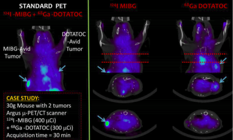
| ABSTRACT | Introduction: Simultaneous in-vivo imaging of several biological processes may improve oncological, neurological and cardiovascular studies by providing complementary information obtained under the same exact conditions and coregistered in space and time. Standard PET imaging does not allow multiplexed acquisitions, as all annihilation photons have the same energy. We developed a technique called multiplexed PET (mPET), which uses a tracer labelled with a pure positron emitter (such as 18 F, 13 N, 11 C), and a tracer labeled with a positrongamma emitter (such as 124 I, 76 Br, 82 Rb, 86 Y). Positrongamma emitters generate a significant number of triplecoincidences , which allows them to be differentiated from the standard PET radionuclides . In this work, we evaluated the performance of mPET using phantom and animal experiments. Methods: The acquisition software of the preclinical Argus PET/CT scanner was adapted to provide datasets containing either double or triple coincidences. Double coincidences were reconstructed using the standard MLEM algorithm, and the triples using a modified version of MLEM, in which each triple is represented by a Vshaped line of response. The separated images of each radiotracer were obtained with an iterative algorithm based on the calibrated sensitivity of detecting double and triple coincidences for each radionuclide. The Image quality provided by mPET was evaluated using a NEMA IQ phantom filled with either 18 F, 124 I or both and by animal experiments (with 68 GaDOTATOC and 124 IMIBG). Separated images of each tracer obtained with mPET were compared in all cases with equivalent singleradionuclide PET images.Results: mPET provided images with little bias (<10%), similar spatial resolution and contrast, and a small relative noise increase (<10%) compared with that obtained with single tracer PET acquisitions.Conclusions: These results show the feasibility of the mPET technique in realistic preclinical scenarios. As it may be used with other PET scanners, it holds the potential for improving the capabilities of molecular imaging.Acknowledgement: This work was supported in part by Comunidad de Madrid (Spain) through the Madrid –MIT M+Visión Consortium, the COFUND of the Seventh Framework Program of the European Union, and the Human Frontier Science Program grant RGP0004/2013. |
| LINK | Link |