| AUTHORS | |
| JOURNAL | ARVO |
| ABSTRACT |
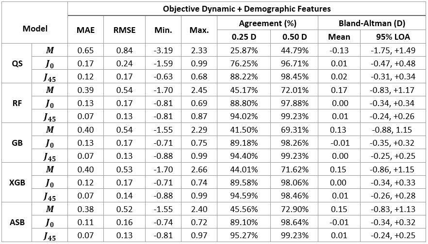
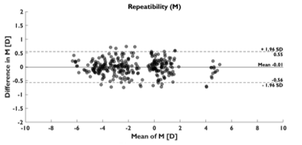
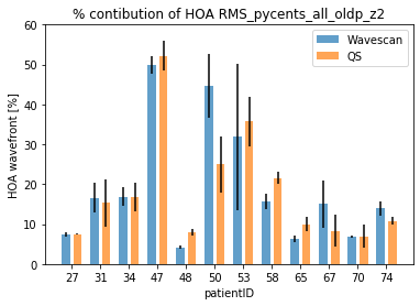
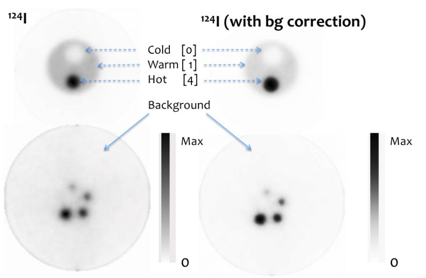
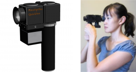
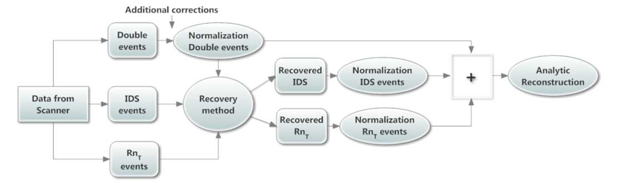
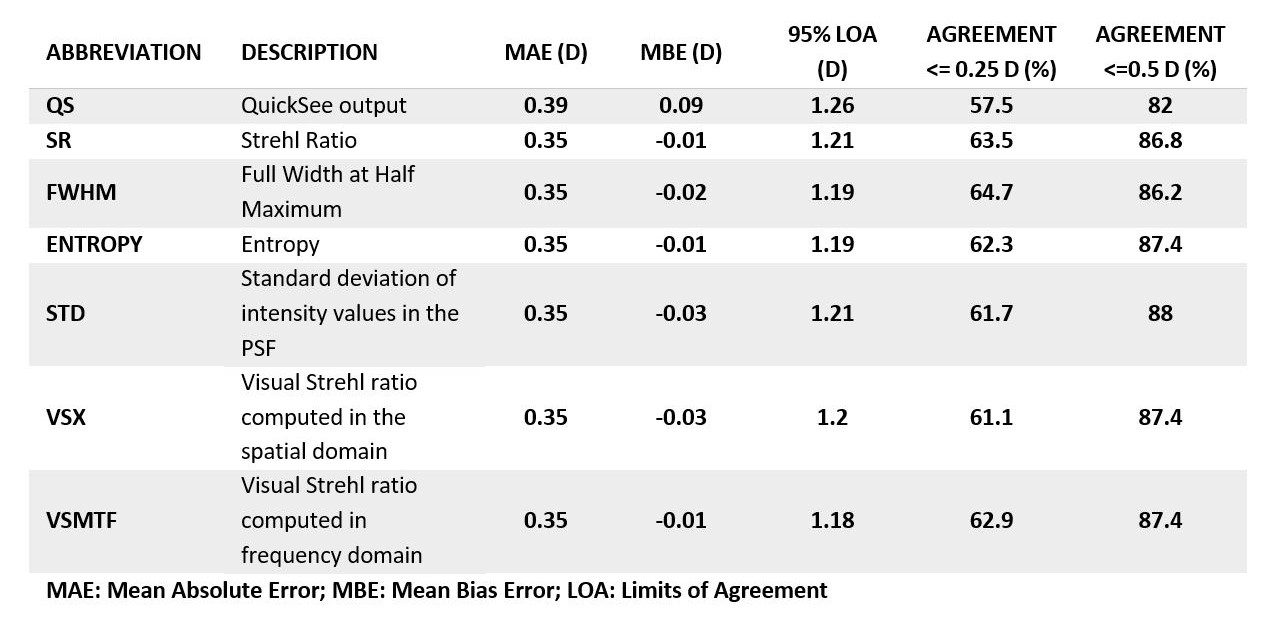
PurposeModern autorefractors have demonstrated high accuracy and higher repeatability than manifest refraction in adults yet are not considered precise enough to substitute the gold standard, even in low-resource settings where there is a severe shortage of experienced refractionists. This work evaluates a novel approach that combines dynamic wavefront aberrometry data, acquired using an affordable portable autorefractor, with the analysis of retinal image quality metrics (IQMs) to predict subjective refraction. Methods56 subjects (34±14 years) were recruited for the analysis. Each participant underwent standard clinical refraction followed by a 10-second video acquisition using a QuickSee (QS) wavefront aberrometer (PlenOptika, USA). Shack-Hartmann images of each video for the right eye were processed to obtain Zernike coefficients up to the 4th order. Coefficients obtained for each image were mathematically corrected with the closest sphero-cylindrical correction and the residual wavefront error was used to calculate the Point Spread Function (PSF) and IQMs (e.g., Strehl Ratio) describing some performance parameter of the corrected eye. Since each IQM is part of a dynamic sequence, it is possible to build a dynamic signal for each metric which contains information about fluctuations in image quality during the measurement. The final refraction is obtained as the average of the refractions corresponding to the images whose IMQs provided optimal performance. ResultsThe proposed method reduced the differences between QS and manifest refraction. Specifically, for spherical equivalent refraction (M) (Table 1) a 79% average reduction in MBE together with moderate average improvements for MAE (10.3%), LOA (5%), and percentage of agreement within 0.25D (8.8%) and 0.5D (6.3%) were found for all IQMs evaluated. ConclusionsThe proposed algorithm behaves as an efficient filter which selects those measurements within the dynamic sequence that are more representative of the manifest refraction of the patient. |
| LINK | here |