Multiplexed PET in Clinical Scanners Based on Triple Coincidences
| AUTHORS | |
| JOURNAL | IEEE Nuclear Science Symposium & Medical Imaging Conference, 2014 |
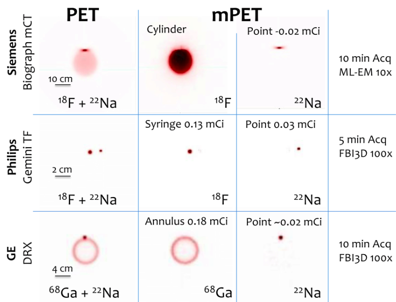
| ABSTRACT | Simultaneous invivo imaging of the biodistribution of two or more radiotracers of interest with Positron Emission Tomography (PET) could provide more comprehensive information about specific targets and physiological processes of interest; however, conventional PET cannot separate different positronemitting radionuclides based on the energy of the detected gamma photons. We have shown previously that the detection of triplecoincidence events allows the separation of radiotracers labeled with positron + gamma emitters (like 124 I) from others labeled with standard pure positron emitters (like 18 F), without requiring any kinetic modelling or precise energy measurement. The goal of the current study was to demonstrate that this methodology can also be applied to clinical PET scanners. On three different clinical PET/CT scanners (GE DRX, Siemens Biograph mCT, and Philips Gemini TF), we recovered triple coincidence events from standard list mode events that were stored as consecutive double coincidences. Phantoms containing either 18 F + 22 Na or 68 Ge + 22 Na were imaged. For all scanners, the separated images of the two simultaneously acquired radionuclides were quantitatively equivalent (bias values below 5%) to those reconstructed from separate acquisitions, with acceptable (below 5%) increase in image noise. For 22 Na, the percentage of detected triple coincidence events ranged from 1.3 to 2.4 % of the doubles for the three systems studied. These results are the first experimental demonstration of simultaneous dualradionuclide PET imaging in commercial scanners in static acquisitions. They were obtained without requiring any software or hardware modification of the scanners. |
| LINK | Link |