A SPECT Scanner for Rodent Imaging Based on Small-Area Gamma Cameras
| AUTHORS | |
| JOURNAL | IEEE TRANSACTIONS ON NUCLEAR SCIENCE, VOL. 57, NO. 5, OCTOBER 2010 |
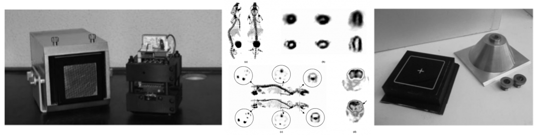
| ABSTRACT | We developed a cost-effective SPECT scanner prototype (rSPECT) for in vivo imaging of rodents based on small-area gamma cameras. Each detector consists of a position-sensitive photomultiplier tube (PS-PMT) coupled to a 30 x 30 Nal(Tl) scintillator array and electronics attached to the PS-PMT sockets for adapting the detector signals to an in-house developed data acquisition system. The detector components are enclosed in a lead-shielded case with a receptacle to insert the collimators. System performance was assessed using 99mTc for a high-resolution parallel-hole collimator, and for a 0.75-mm pinhole collimator with a 60° aperture angle and a 42-mm collimator length. The energy resolution is about 10.7% of the photopeak energy. The overall system sensitivity is about 3 cps/μCi/detector and planar spatial resolution ranges from 2.4 mm at 1 cm source-to-collimator distance to 4.1 mm at 4.5 cm with parallel-hole collimators. With pinhole collimators planar spatial resolution ranges from 1.2 mm at 1 cm source-to-collimator distance to 2.4 mm at 4.5 cm; sensitivity at these distances ranges from 2.8 to 0.5 cps/μCi/detector. Tomographic hot-rod phantom images are presented together with images of bone, myocardium and brain of living rodents to demonstrate the feasibility of preclinical small-animal studies with the rSPECT. |
| LINK | http://ieeexplore.ieee.org/document/5571005/ |