| ABSTRACT |
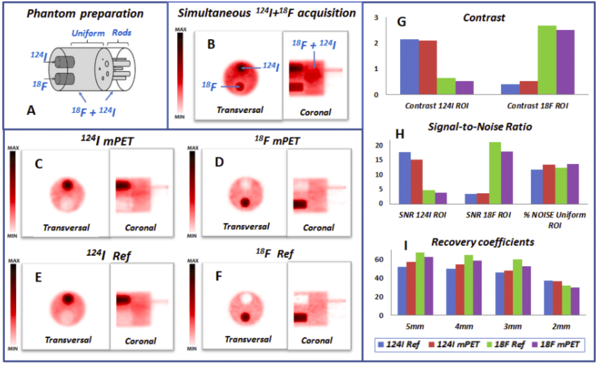
Positron emission tomography (PET) is one of the most sensitive noninvasive molecular imaging tool, being its sensitivity several orders of magnitude higher than that typically obtained in single photon emission computed tomography (SPECT). However, PET lacks the ability of SPECT to multiplex signals from several tracers, which is very useful in many different studies such as cardiac imaging with 99mTcSestamibi and 201Tl. Recently, it has been shown that the use of tracers labelled with positrongamma emitter radionuclides like (124I, 86Y, 82Rb, 94mTc, 76Br) in combination with tracers labelled with standard positronemitter radionuclides like (18F, 11C, 13N) enables multiplexed PET (mPET). mPET uses the triple coincidences from the positrongamma emitters, together with the standard double coincidences to reconstruct separated images of each radionuclide’s activity distribution. We obtained encouraging results with mPET in some initial preclinical studies, but a detailed study of the quality and quantification properties of mPET images, and an evaluation of its performance in realistic clinical scenarios was still required.
|